Stem Cell Technologies to Integrate Bio Design-Related Tissue Engineering within the Frame of Cell-Based Regenerative Medicine: Towards the Preventive, Therapeutic and Rehabilitative Resources and Benefits
1. Editorial
The ultimate goal for regenerative medicine is to channel
multipotent human cells with high proliferative capacity into pre-defined
scenarios and specified differentiation programs within the human body.
Meanwhile, end-stage heart failure is a global
scourge, and cardiovascular disease (CVD) is a major health problem and the
leading cause of morbidity and mortality in the Globe. And thus, the treatment
and prevention of CVD are considered to be the public health priorities. The
latter means that CVD is a life course disease that begins with the evolution
of risk factors that in turn contribute to the development of subclinical
(pre-early and symptom-free) atherosclerosis.
The idea of extending the lifetime of the human heart
has been fuelled by a series of major advances in transplantation and drug therapies.
Nevertheless, atherosclerotic complications and myocardial infarction, in
particular, is characterized by the irreversible loss of cardiac myocytes
because of the ischemic necrosis. Therefore, the need to re-establish the
structural and functional features of native heart tissue represents a major
challenge for the field of cardiac regeneration and engineering as the latest
avenue to move ahead.
Stem Cell (SC)-based therapy has been considered as a
promising alternative in the treatment of ischemic heart disease. And Cardiac
Stem Cells (CTCs) are described in a number of mammalian species including
humans, and their clusters are considered to consist of both lineage-negative
and partially committed cells which, in turn, are located among contracting
cardiac myocytes. CSCs are directly involved in cardiac cellular homeostasis
during aging and adaptation to physiological and pathological stress and being
transplanted into damaged hearts, CSCs have the capacity to generate de novo
myocardial tissue. The niche for CSCs can be activated by several active
biomolecules (including cytokines and growth factors), or through the injection
of systemic drugs, such as statins, to obtain beneficial results similar to
those of CSC transplantation.
Meanwhile, current understanding defines the heart as an organ comprised of heterogeneous population of myocytes, which continue to die and self-renew, thereby maintaining cardiac integrity throughout the life. However, the cell source for heart self-renewal, purportedly by the replacement of senescent cardiac myocytes (CMs) with juvenile cells, as well as the underlying mechanisms remain obscure (Figure 1).

Figure 1: Potential cell sources for heart regeneration therapy.
Meanwhile, dedifferentiation of adult CMs, trans
differentiation of endogenous stem cells (SCs), and fusion of SCs with cells of
other types are considered as major and event customized scenarios whereby new
CMs are generated in adult heart.
But although SC administration resulted in the temporary improvement of myocardial contractility, the SC-induced formation of new cardiac myocytes within the injured part of the heart (myocardium) has not been proven. So far, current therapies merely delay its inexorable progression, and the lack of a clinically-suitable and highly productive human Cardiac Myocyte (CMC) source with proper myocardium regenerative potential has been the major setback in regenerating the damaged human heart, either by endogenous cells or by cell-based transplantation or cardiac tissue remodelling and engineering via procedures of bio- and/or drug design being based on drug discoveries. The latter does strongly need much more advanced and promising techniques to improve morphological and electromechanical properties of the diseased heart (Figures 2-4).

Figure 2: Stem cell-based strategies for cardiac regeneration after heart disease.
Multiple techniques to improve morphological and
electromechanical properties of the diseased heart: (1) In vitro cardiac
differentiation of different stem cell types. (2) Tissue engineering approaches
combining cells with biomaterials to design in vitro cardiac patches or
injectable scaffolds for transplantation into the infarcted heart area. (3)
Cell-and gene-based strategies secreting cytokines, growth factors and
microRNAs to promote cardiac regeneration. (4) Stem-cell derived exosomes as an
innovative cell-free therapy in heart regenerative medicine.
Right now, different cell types are under evaluation
regarding their regenerative potential. First-generation cell types including Skeletal
Myoblasts (SMs), Bone Marrow Mononuclear Cells (BMMNCs), Hematopoietic Stem
Cells (HSCs), Endothelial Progenitor Cells (EPCs), and Mesenchymal Stem Cells (MSCs)
were initially introduced. Despite promising preclinical studies,
first-generation approaches displayed heterogeneous clinical outcomes.
Variations between trials may be attributed to differences in design and the efficacy of the first-generation cell-based approaches and came to divergent conclusions. Nevertheless, the field partially switched to second-generation cell types including lineage-guided cardiopoietic cells, cardiac stem/progenitor cells (CSCs/CPCs), and pluripotent stem cells (Figure 3).

Figure 3: Evolution of translational cardiac regenerative therapies.
First-generation cell types such as SMs, BMMNCs, HSCs, EPCs, and MSCs demonstrated feasibility and safety with, however, heterogeneous outcomes and limited efficacy in the clinical setting. In order to better match the target organ, second-generation cell therapies propose the use of cpMSCs, CSCs/CPCs, and CDCs, and pluripotent stem cells such as ESCs and iPSCs. Next-generation therapies for cardiac repair are directed toward cell enhancement (e.g., biomaterials, 3D cell constructs, cytokines, miRNAs) and cell-free concepts (e.g., growth factors, non-coding RNAs, extracellular vesicles, and direct reprograming)

Figure 4: Cell therapy goals in cardiovascular disease.
1.1. The CSC's main role is to replace myocardial
dying CMCs and reconstruct damaged areas in the heart
CMCs mentioned above contribute to most of the structural volume of the heart. The relative simplicity in development and maturation of the embryonic heart makes it also possible to be the first organ to be reconstituted, for instance, from human embryonic stem cells (hESCs) (Figure 5).

Figure 5: Human embryonic stem cells derivation and characteristics.
Establishing a controllable differentiation route to
efficiently generate a large supply of human cardiac elements from hESCs
cardiac derivatives will make it become feasible to reconstruct the human 3D
beating heart that would reflect the biological complexity and microenvironment
niche of the in vivo human heart and function, and will facilitate progress in
the identification of potential therapeutic targets for prevention and
treatment of CVD. Globally, the latter will provide ground-breaking technology
platform for tissue reconstitution from hESC-derived somatic elements,
innovating in regenerative medicine that will have a tremendous impact on
translational medicine, daily clinical practice, the healthcare biopharma and
industry, and the market of the next-step generation, finally.
In reality, in the adult heart, the mature contracting CMCs are terminally differentiated and unable to regenerate. So, damaged or diseased CMCs are removed largely by macrophages and replaced by stromal (scar) tissue. Although stem/progenitor cells have been identified in postnatal hearts, the minuscule quantities and growing evidences indicating that they are not genuine heart cells have caused skepticism as being harnessed for cardiac repair.
2.
SC-derived paracrine factors the newest effective tools to repair the
myocardial damages and to manage cardiac healthy entity
The development of the heart appears to be regulated
by complex signalling networks and proven to be generated from two distinct
progenitor cell populations or heart fields that segregate from a common
progenitor at gastrulation. The primary and second fields are regulated and can
be distinguished by the expression of a broad spectrum of humoral biomolecules
including specific transcription factors and signalling molecules. And
developing cellular models of human embryonic heart formation will reveal the
biological pathways and molecular targets that control cardio myogenesis,
thereby, aids identification of therapeutic targets for the prevention and
treatment of heart disease and failure.
So, some focus of basic research in the field and translational applications have since shifted to SC-derived paracrine factors, including cytokines, growth factors, mRNA, and miRNA (notably, the latter can enter into the extracellular space either in soluble form or packed into membrane vesicles) (Figure 6).

Figure 6: Cardiac regenerative strategies via paracrine mechanisms.
Schematic representation of the main experimental
cardiac medicine approaches suggested to address myocardial injury and aiming
at stimulating endogenous mechanisms of repair and myocardial restoration by
means of stem cell-derived paracrine effectors and biomaterials.
And then the paracrine factors have been shown to
suppress inflammation and apoptosis, to stimulate angiogenesis, and to induce
and amplify the proliferation and differentiation of resident cardiac SCs
(CSCs). Such features have led to exosomes being considered as potential drug
candidates affording myocardial regeneration. The search for chemical signals
capable of stimulating cardiomyogenesis is still ongoing despite continuous
debates regarding the ability of mature cardiac myocytes to divide or to
dedifferentiate, and the ability of CSCs to differentiate into cardiac
myocytes.
In this sense, one of the most effective tools to manage cardiac entity and to control the balance of CMCs within the myocardium whilst preventing subclinical stages of the real-time disorder and the disease, to treat the latter and to regenerate the post-treatment complications and damages could be SCs and biomolecules secreting by those cells and cells in the SC microenvironment. Among families of SCs are the heart muscle cells (cardiac myocytes/CMCs) being specific for congenital and chronic inflammatory heart cases, ischemic disorders or for heart attack victims. For instance, human embryonic stem cells (ESCs) and induced pluripotent stem cells (iPPSCs) can self-renew indefinitely, while maintaining the capacity to differentiate into adult and useful CMCs. So, the increasing availability of human iPPSCs provides new prospects for the cell replacement strategies and disease-related basic research and translational applications. So, the availability of human stem/progenitor/precursor cells and SC/progenitor cell inducers and activators in high purity with adequate cardiogenic potential will greatly facilitate developing safe and effective cell-based regeneration and replacement therapies against heart disorders. It makes heart disease and failure possible to be the first major health problem to be resolved by clinical translation of the advances of hESC research and applications.
2.1. Pluripotent stem cells (PPSCs): the first steps
towards the cardiologist’s practice
The ongoing rapid technological advances in hiPSC translational research have been directed toward the clinical application of this technology. Among broad populations of SCs, BM-derived and cardiac-specific SCs have demonstrated potential for cardiac regeneration. And no secrets that PPSCs (Figure 7).

Figure 7: Pluripotent stem cells (PPSCs) on the way to generate cardiac myocytes.
With their ability to self-renew and differentiate into every cell type of the body, have attracted significant interest for understanding basic biology and the development of translational applications.
Embryonic Stem Cells (ESCs) and induced Pluripotent
Stem Cells (iPSCs), which are collectively called PPSCs, have emerged as a
promising source for regenerative medicine and cardiology. Particularly, human
pluripotent stem cell-derived cardiomyocytes (hPSC-CMCs) have shown robust
potential for regenerating injured heart, opening the door for clinical
application to secure the cardiac repair.
Cell therapy with hPSC-CMCs has shown great potential for individualized therapy of injured heart, is expected to serve as an integral component of PPM and is potentially viewed as a treatment that would revolutionize the management of patients with severe heart failure (Figure 8).

Figure 8: Strategy of cardiac regenerative therapy using hIPSC-derived CMCs.
However, more studies are needed to ensure the precise
therapeutic effects, underlying mechanisms, and safety, before this technology
can be applied clinically.
2.2. Induced Pluripotent Stem Cells (iPSCs)
A novel approach in cardiac regeneration has been
proposed with the discovery of induced pluripotent stem cells (iPSCs).
iPSCs nearly identically resemble ESCs and can give
rise to all cell types (including CMCs) in the body, and thus have opened new
opportunities for PPM and new ways of modeling human diseases. iPSCs are able
to efficiently differentiate into CMCs and thus hold a real regenerative
potential for future clinical applications.
Better understanding and control of the reprogramming
process should enable enhanced efficiency and higher fidelity in reprogramming.
Toward those trend and path, the possibility to directly induce conversion of
fibroblasts into CMCs has recently emerged as a promising area for in situ
cardiac regeneration. And better understanding and control of the reprogramming
process should enable enhanced efficiency and higher fidelity in reprogramming.
iPSCs which are reprogrammed from somatic cells, allow
for the generation of patient-specific (personalized) pluripotent cells
(PSPPSs) and have diminished ethical concerns, and so are promising for
personalized disease modeling (to secure matching a patient to a treatment) and
regenerative medicine. Over the past few years, various combinations of
biomolecules (including transcription factors/TFs), have successfully been
developed to create iPSCs. Inspired by the iPSC approach using multiple TFs,
many studies have shown that, with the proper conditions, somatic cells can
also be transdifferentiated into another cell fate both within and outside of
their germ layer, which is also called lineage-specific reprogramming.
In particular, CMCs differentiated from iPSC may represent a realistic option for the regeneration of the injured heart. A significant number of different cell therapy strategies that employ iPSC-CMs have been proposed to restore cardiac function, ranging from injection of cells alone, to their combination with ECM-like biomaterials, and to more complicated engineered-based strategies, which aim to faithfully recreate the myocardium. The majority of these approaches have shown - at least to some extent - success in mimicking heart cells and improving cardiac function, providing new hope for the replacement of CMCs after the irreversible loss of heart tissue occurring during myocardial infarction (Figure 9).

Figure 9: hPSC-based bioengineering strategies for cardiac regeneration.
The panel (A) shows a schematic representation of the
different hPSC-based methodologies used for regenerative purposes in the
cardiac field. The panel (B) provides a time line that summarizes the key
milestones reached in the field, starting from the simple injection of hPSC-CMs
into the heart to the development of tissue-like structures with enhanced
hPSC-CM maturation, more complex perfusable and personalized constructs and
injectable hydrogels.
Notably, Menasché et al. reported
the first clinical application of cardiac patches composed of human ESC-derived
cardiac progenitor cells in a patient suffering from severe ischemic left
ventricle dysfunction, demonstrating the possibility to use a engineered-based
approaches for cardiac repair.
2.3. Bone fide CSCs
Meanwhile, the discovery in the adult heart of bone
fide CSCs put the first brick into a new field of cardiac regenerative biology
and medicine that has deeply changed the outlook of the potential for treating
cardiac failure.
2.4. Mesenchymal stem cell-based drugs and their
applications in cardiology
Along with the introductory SC-related reference, the stroma-related cells, or mesenchymal stem cells (MSCs) (Figure 10), being multipotent progenitor cells would constitute a minute proportion of Soluble factors released by MSC play an essential role in the post-ischemic reparative process improving angiogenesis, cytoprotection, and endogenous cardiac regeneration and reducing fibrosis. Ang-1 angiopoietin 1, HGF hepatocyte growth factor, MSC mesenchymal stem/stromal cells, VEGF vascular endothelial growth factor the bone marrow, represented as a rare population of cells that makes up 0.001 to 0.01% of the total nucleated cells. MSCs are able to differentiate into both mesenchymal, as well as, non-mesenchymal cell lineages, such as myocytes, both in vitro and in vivo. In this sense, several studies have documented the substantial clinical improvements observed in animal models, when MSC were systemically introduced as a therapy in mouse models of myocardial ischemia and infarct.

Figure 10: MSC paracrine action/mechanisms in heart regeneration.
As described previously, MSCs are characterized by their hypoimmunogenicity which would secure allo-MSC engraftment in cardiac tissues in the absence of immunosuppression, whilst favourably altering ventricular function. The allo-MSC engraftment occurred without evidence of immunologic rejection and in the absence of assisted immunosuppressive therapy emphasizing some of the apparent advantages of those cells over other cell populations for cellular cardiomyoplasty (Figure 11).

Figure 11: Cellular cardiomyoplasty as a new translational and therapeutic approach involving engineering culture systems, the use of novel biomaterials for mechanical support of the cells and for controlled release of therapeutics, and tissue engineering.
Diagrammatic representation of the different
approaches that can be used to repair infarcted myocardial tissue whilst securing
the cellular cardiomyoplasty. An acellular patch can be used as an
off-the-shelf product that can be implanted soon after myocardial infarction.
Alternatively, cells can be harvested (i.e., progenitor cells) and injected
back into the patient. Another approach can be the isolation of somatic cells
(i.e., blood cells), reprogrammed, expanded, differentiated, and assembled into
a bioengineered cardiac tissue that can then be implanted back into the patient
as an autologous patch. These approaches have different timing and expenses
associated with them that can have potential impact on their clinical use.
The concept of adult SC plasticity [Wagers AJ,
Weissman IL. Plasticity of adult stem cells. Cell. 2004;116: 639–648.] implies
that SCs and CSCs can transdifferentiate into mature cell types outside their
original lineage in response to microenvironmental cues. For example, HSCs may
transdifferentiate into CMCs, thereby improving heart function and survival
[Orlic D, Kajstura J, Chimenti S, Jakoniuk I, Anderson SM, Li B, Pickel J,
McKay R, Nadal-Ginard B, Bodine DM, Leri A, Anversa P. Bone marrow cells
regenerate infarcted myocardium. Nature. 2001;410: 701–705.]. Meanwhile, SCs
release angiogenic ligands, protect cardiomyocytes from apoptotic cell death,
induce proliferation of endogenous CMCs, and may recruit resident CSCs.
As a whole, the data accumulated from preclinical and
clinical data indicate that bone marrow-derived MSCs have, in addition to their
therapeutic purposes in regenerative medicine, effects that can result from
their anti-inflammatory properties. In addition, the therapeutic effectiveness
of MSCs relies heavily on their ability to modify microenvironments. Those
modifications occur through the release of cytokines, and anti-apoptotic and trophic
molecules that promote the repair and protection of damaged (including cardiac)
tissues.
2.5. Human embryonic stem cell (hESC)-based drugs
Currently, the hESC CMC therapy derivatives are the only available human cell sources with adequate capacity to regenerate contractile heart muscles, vital for heart repair in the clinical setting (Figure 12).

Figure 12: Approaches to preparing hESC-derived cardiac myocytes for tissue repair.
Researchers are focusing on chemical (e.g., 5-azacytidine and p38 MAPK inhibitors) and biological (e.g., activin A, bone morphogenetic protein, basic FGF, VEGF and Dickkopf homolog 1) factors, genetic (e.g., miRNAs) and epigenetic (e.g., miRNAs and chromatin remodeling) manipulation, and mechanical factors (e.g., hydrodynamics and surface tension) to direct cardiomyocyte differentiation from hESCs. These approaches are complemented by purification methods that take advantage of the biochemical properties of human cardiomyocytes (e.g., Percoll density centrifugation and mitochondrial content), and selection strategies that rely on the expression of cardiac-specific genes (e.g., reporter lines and molecular beacons) and surface markers. hESC: Human embryonic stem cell.
Directed differentiation of hESCs has generated much
interest in the field of regenerative medicine. Because of their ability to
differentiate into any cell type in the body, hESCs offer a novel therapeutic
paradigm for myocardial repair by furnishing a supply of CMCs that would
ultimately restore normal myocardial function when delivered to the damaged
heart. Moreover, immunosuppressive regimens for hESC-based therapeutics may not
need to be as rigorous as conventional organ transplantation. Nevertheless,
transplanted hESC-derived CMs will be susceptible to immune rejection to some
degree.
Spontaneous CMC differentiation of hESCs is an
inefficient process that yields very low numbers of CMCs, the need for new
methods of directed differentiation of hESCs into functional CMCs and cardiac
progenitors has led to an explosion of research utilizing translational
strategies to direct cardiac differentiation and enrich populations of cardiac
cells for therapeutic use. Further improving policy making and funding
situation for hESC research and translational applications would open up a new
dimension of cell therapy-based future medicine to provide new treatments for
life-threatening cardiovascular diseases and failures. Transforming pluripotent
hESCs into fate-restricted therapy derivatives would dramatically increase the
clinical efficacy and safety of hESC-derived cellular products, bringing
cell-based regenerative medicine to a turning point.
Meanwhile, the discovery that somatic cells can be reprogrammed to iPSCs has attracted enormous interest in translational research and applications. Therefore, small biomolecules regulating reprogramming mechanisms are valuable tools to probe the process of reprograming and harness cell fate transitions for clinical applications (Figure 13).

Figure 13: Schematic representation of the workflow for iPSC generation and differentiation from patients’ somatic cells and major applications to human health.
As you would see, engineering approaches toward the repair of myocardial tissue have shown promise in the past, but full restoration of myocardial function remains elusive. As our understanding of the heart's physiology and function grows, we would suggest the potential approaches as: (1) acellular scaffolds that provide bioactivity and biomechanical support to the heart and (2) cellular scaffolds that provide the minimum combination of cells and biomaterial composition needed for increased bioactivity and support. It is also clear that advanced culture systems will be needed to create more advance engineered cardiac tissues and to create better screening tools to test bioengineered cardiac patches in vitro.
Anyway, the heart has an endogenous myocardial regenerative potential owed to CSCs. But, to date, the lack of a clinically-suitable source of engraftable human stem/progenitor cells with adequate cardiomyogenic potential and the appropriate engineered cellular scaffolds that have been the major setback in developing safe and effective cell-based therapies for regenerating the damaged or lost cardiac muscle structure and circuitry in a wide range of cardiac disorders and damaged human heart. Moreover, current cell delivery methods to the damaged heart, by injection of cells either directly into the infarcted region or via the coronary circulation, are inefficient. In addition, arrhythmogenesis is a potential risk in cell-based cardiac repair. So, future research is aimed at identifying novel cell type candidates being capable of differentiating into cardiac myocytes. The observation that CSCs can undergo intracellular development with the outcome formation of “cell-in-cell structure” and subsequent release of transitory amplifying cells with the capacity to differentiate into cardiac myocytes may provide clues for stimulating regenerative cardiomyogenesis.
3.
Understanding SC-based therapy of the second-step generation: the novel cell
type candidates to secure regenerative cardiomyogenesis
SCs and CSCs, as subjects to be utilized in treating
cardiovascular diseases, are envisioned as a replacement for lost functional
cells (e.g., CMCs), a means of trophic support, and, more recently, a
cell-based therapeutic tool for in vitro modelling to understand disease and to screen and
personalize treatments. The need for new and improved pharmacotherapies in
modern cardiology to treat and, moreover, to prevent the disease while
minimizing preclinical manifestations is driving interest by the specialists in
personalized and Precision Medicine (PPM).
Consistent with the traditional view that the heart is a “postmitotic” organ that possesses minimal capacity for self-repair, much of the preclinical and clinical work has focused exclusively on introducing SCs into the heart, with the hope of differentiation of these cells into functioning CMCs. This approach is ongoing and retains promise but to ate has yielded inconsistent successes. More recently, it has become widely appreciated that the heart possesses endogenous repair mechanisms (Figure 14) that, if adequately stimulated, might regenerate damaged cardiac tissue from in situ cardiac stem cells.

Figure 14:
Endogenous cardiac regeneration.
After a cardiac insult, a significant number of CMCs
die and vessel density gets reduced. In a very limited way, the heart has the
ability to regenerate but it is insufficient to compensate for the total
damage. The cellular participants in the endogenous regeneration process may
include CMCs (pink cells, black lines), local cardiac progenitor cells (blue
cells), and recruited peripheral stem cells (red cells). These cells have the
potential to proliferate and participate in the regeneration of CMCs, angiogenesis,
and the release of trophic factors that may reduce cardiac cell death. Modes of
the cell’s participation are identified by colored arrows (peripheral stem
cell, red; cardiac progenitor cell, blue; cardiac myocyte, green). Methods that
could be employed to improve these endogenous mechanisms include cell and/or
gene therapy and pharmacologic treatments.
Accordingly, much recent work has focused on engaging
and enhancing endogenous cardiac repair mechanisms. So, understanding the
biological activity of SCs, CSCs and progenitor cells, and their ability to
contribute to the repair, regeneration and remodeling of the human heart is
just crucially important.
Among the latest SC-based products is Baxter
Healthcare’s Phase III trial of
intramyocardially administered autologous HSCs (harvested from peripheral
blood), intended to be used in patients with refractory chronic myocardial
ischemia. Findings to date indicate that the product can repair heart tissue,
increase blood flow, and allow the patient to exercise.
One direct competitor to Phase III trials Baxter’s
treatment is AMR-001 (NeoStem), which uses autologous enriched HSCs from the
bone marrow to treat ST elevation myocardial infarction. Injected into the
infarct-related artery, AMR-001 targets the site of ischemic injury, where
chemokine CXC receptor 4 (CXCR4) on HSCs binds to stromal-derived factor 1
(SDF1), which is induced by hypoxia-inducible factor (HIF) produced by ischemic
tissueOther late-stage candidates include Aastrom’s Ixmyelocel‑T (Figure 15).

Figure 15: Production of ixmyelocel-T and its applications in cardiologist’s practice.
Ixmyelocel-T is composed of a mixture of cell types
that include those expected to be found in the BM-MNC population. These include
myeloid cells (granulocytes, monocytes, and mixed myeloid progenitors) and
lymphoid cells (T cells, B cells, and mixed lymphoid progenitors) that express
CD45 on the cell surface, CD90+ MSCs, and CD45+CD14+ autofluorescent+
(CD14+Auto+) macrophages. The numbers of CD90+ and CD14+Auto+ cells are
significantly greater in ixmyelocel-T because of expansion during the Aastrom
(now Vericel) proprietary expansion process.
which consists of autologous cells derived from BM and
proved to promote immunomodulation, angiogenesis and tissue remodeling. The
product is in Phase III trials for critical limb
ischemia and dilated cardiomyopathy.
Bioheart and Cytori also have Phase III C‑Cure (Cardio3 Bioscience) which is based on
autologous MSCs differentiated via its cardiopoiesis platform into CMCs, which
are re-injected into the heart. Cytori’s adipose-derived MSCs for acute
myocardial infarction underwent Phase II/III trials.
Evidence has been presented that a fraction of CMCs
may be able to reenter the cell-cycle and that limited regeneration can occur
through recruitment of resident and circulating SCs. Some clinicians may regard
these new ideas as being mere curiosities, because of their everyday experience
that endogenous repair mechanisms are overwhelmed in patients with acute
myocardial infarction (AMI), advanced coronary artery disease, and chronic
heart failure. However, the existence of endogenous repair mechanisms suggests
that cardiac repair may be achieved therapeutically in these clinical settings.
The occurrence of viable cells internalized within different kinds of host cells has been recognized for more than 100 years and usually to illustrate: (i) cannibalism, (ii) emperipolesis and (iii) and entosis, which differ in both effector and host cell identity, mechanism of penetration, and function. Both emperipolesis and entosis appear to share similar features with the development of intracellularly localized CSCs which are being encapsulated are able to replicate followed by the partial cardiomyogenic differentiation (Figure 16-18).

Figure 16: The CSCs inside CMCs and the formation of CSC-containing CICSs in the cultures obtained from newborn and 20- and 40-day-old rats.
(A) Experimental design. The cells were plated and
cultured for up to 30 days, followed by immunostaining or time-lapse
microscopy. (B–G) Immunocytochemistry. The nuclei of the cells have been
stained with Hoechst. Transmitted light and fluorescent images are merged. (B)
c-kitC CSC inside a CM obtained from a newborn rat (day in vitro 6). (C) Isl1C
CSC inside a CM obtained from a newborn rat (day in vitro 4). As documented by
the expression of Ki67, both the CSC and the host cell exhibit proliferative
ability. (D) ScaC CSC encapsulated between the nuclei of the host cell
(20-day-old rat, day in vitro 11). (E) A mature c-kitC CSC-containing CICS with
a prominent coating (“capsule”) with 3 pores (white arrows, 40-day-old rat, day
in vitro 6). Optical sectioning
shows the host cell nucleus (blue) just above the CICS (see sections 13–15 in Video S1). (F-G) The CICS capsule in detail. (F) Erosion of the Isl1C CSC-containing CICS capsule (black arrow) obtained from a 40-day-old rat, day in vitro 8. The pores are also visualized (white arrows). The capsule interior is positive for sarcomeric a-actinin, also observed in (G). (G) Erosion of the c-kitC CSC-containing CICS capsule (black arrow) obtained from a newborn rat, day in vitro 20. The pores are seen (white arrows).

Figure 17:
Optical tomography of the CSC-containing CICSs.
The optical sections were spaced 1.01 mm (A, B) and 2.01 mm along the z-axis (C). Images are placed in consecutive order from the bottom to the top of structures. (A) C-kitC CSC-containing CICS in the culture obtained from a 40-day old rat (FITC, green, day in vitro 6) After counterstaining for a-sarcomeric actin (Alexa 543 nm, red), the nuclei (Hoechst, blue) of both the CSC and the host cardiomyocytes are visualized; the vertical dimension of the CICS is 20 mm (slices 3 to 20). Transmitted light and fluorescent images are merged. (B) The Isl1C CSCcontaining CICS (FITC, green, left column) inside a given host CM obtained from a newborn rat (day in vitro 11), with a vertical dimension of 7 mm (slices 2 to 6). Cytoskeletal actin (rhodamine phalloidine, red, central column) can be seen. Green and red fluorescent images are merged and presented in the right column. (C) The Isl1C CSC-containing CICS in the culture of newborn rat (day in vitro 4). Isl1C CSCs (FITC, green), cytoskeletal actin (rhodamine phalloidine, red), the nuclei (Hoechst, blue) are merged with transmitted light images (Figure 18).

Figure 18: Cell-in-cell structures (CICSs) identified in the suspension of freshly isolated myocardial cells (ex vivo) of 20- and 40-day-old rats.
Transmitted light and fluorescent images are merged.
(A and B) Isl1C CSCs inside cardiomyocytes of 20-day-old rats (Isl1, green),
a-Sarcomeric actin, red). (C) c-kitC CICS. (40-day-old rat, c-kit, green; Ki67,
red). (D) c-kitC CICS. (40-day-old rat, c-kit, green; a-Sarcomeric actin, red).
Our results suggest that self-renewal of cells in myocardium is driven primarily by proliferation and differentiation of CSCs inside the colonies and by division and partial differentiation inside the bodies of small myocardial cells (TACs and young CMs). Two processes of mature cardiomyocyte formation are schematically shown in (Figure 19).

Figure 19: Schematic illustration of two pathways whereby mature CMs are generated from CSCs in mammalian myocardium.
The left side of the proposed scheme reflects the process of colony formation, while the right side depicts the stages of intracellular development of CSCs. We propose that the process of colony formation is associated with several rounds of CSC division, eventually resulting in formation of TACs with
The presence of CSC-derived colonies, CICSs and TACs
in myocardium proved our viewpoint about two pathways that generate new CMCs in
adult heart. Moreover, TACs may play a central role in self-renewal of
myocardium throughout the lifetime. By studying the behavior of CSCs, we proved
that the formation of new CMCs from resident CSCs occurs through colony formation
and consequent to their intracellular development inside the CMCs, forming
cell-in-cell structures (CICSs). Our data indicate that cardiomyogenic stimuli
should be focused on TACs, rather than on CSCs or mature CMCs. And the
proliferative activity of TACs is enhanced following ischemia and hypoxia and
that their cardiomyogenic differentiation ability renders them as top
candidates for therapeutic cardiomyogenesis in the diseased heart.
In studies of the last decade [Tyukavin A., Belostotskya G. Zakharov E. et. al., 2015, 2020], it was shown that apoptotic bodies of cardiomyocytes (ApBc) stimulate the development of colonies of resident stem cells of cardiomyocyte precursors in the heart muscle of animals. This is accompanied by an increase in myocardial contractility. The introduction of apoptotic bodies of fibroblasts (ApBf) to experimental animals caused the development of colonies with markers of endothelial cells in combination with a decrease in myocardial contractility (Figure 20).

Figure 20:
Detection of CSC colonies and “cell-in-cell structures” (CICSs) in the
myocardium of “old” rats after exposure to apoptotic cells of cardiomyocytes
(ApBc) and fibroblasts (ApBf).
Earlier on syngeneic animals using the label - GFP was shown [Tyukavin A. et. al, 2012] that a local laser apoptotic effect on tissues causes an intensive transition of mesenchymal bone marrow stem cells (HSCs) from the bloodstream to the zone of programmed cell death (Figure 21).
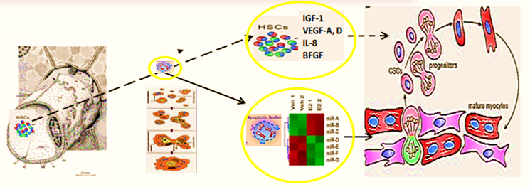
Figure 21: Hypothesis of pathogenesis of restoration of myocardial integrity after injury involving apoptotic bodies of cardiomyocytes (ApBc), resident myocardial stem cells (CSCs), and mesenchymal bone marrow stem cells (HSCs).
Apoptotic bodies (ApB) of cardiomyocytes combine the
functions of CSC and HSCs in the areas of myocardial regeneration. Signaling
molecules are located on the surface of ApB, which mediate the homing and
chemotaxis of HSCs to the area of the damaged myocardium. HSCs provides
targeted delivery of growth factors and cytokines required to maintain CSC
proliferation.
ApB contains a complex of molecules, carriers of
"epigenomic memory" about the tissue belonging of a dead cell. It is
likely that simultaneously with the triggering of the effector link of
apoptosis, which ends with the formation of ApB, RNA is expressed, the new
profile of which is the “code” of the tissue belonging to the dead cell. When
ApB enters the CSC via endocytosis, a specific set of long and short non-coding
RNAs express genes that determine the direction of differentiation of resident
myocardial stem cells. It can be assumed that this hypothesis is true not only
for the heart, but also for other organs and tissues.
Realizing the translational and therapeutic potential
of CICSs has been hindered by some obstacles which would be surmounted and
brought SC-based therapy of the future towards clinical applications, including
establishing defined culture systems for de novo derivation and maintenance of
clinical-grade progenitor cells and lineage-specific differentiation of the
latter by siRNA-driven modulation. Such milestone advances and medical
innovations in CICSs research allow generation of a large supply of clinical-grade
cardiac progenitor-based therapy derivatives targeting for cardiac problems,
bringing cell-based regenerative medicine to a turning point.
4. SC
Therapy and World Market: The future of SC drugs and upgrading niches
The global cell therapy market is continuously
evolving in order to meet the needs of the patients who have been affected by
any particular diseases, and ischemic failure, on particular! The increasing
prevalence of the latter is shifting the market trends from traditional
treatments to novel cell therapies. SC therapies are thus viable alternatives
to conventional treatments with substantial therapeutic potential; market
opportunities are huge, as multiple product candidates are expected to be
approved over the coming decade.
Meanwhile, even with the vast development of the
medical field and surgical interventions, the fact remains that serious
congenital heart diseases (CHDs), being one of the most common disorders in
newborns, are still the leading cause of death and disability for children who
may have to endure long-term problems such as heart failure and other systemic
issues. About 80% of infants and toddlers with CHDs have the potential to reach
adulthood.
At this point, paediatric heart failure is still at its infant phase, and many more studies are needed to elucidate the mechanisms of action. To surmount the barriers, issues such as 3D bioprinting, design-driven gene editing and immune engineering, and the use of organoids for heart tissue modelling are debated on. Steady progress has been realized in treating these diseases using SCs (Figure 22).

Figure 22: PPM-guided regenerative medicine in congenital heart disease.
Advances in regenerative medicine, specifically SC
therapies, have significantly progressed in treating CHD. Different SCs-related
methods are planned to treat pediatric heart problems, thus reducing
complications and improving the heart’s overall function. With advancements
made possible through stem-cell therapy, tissue engineering, and genetic
engineering, avenues have been opened that would lead to developing more
effective treatments for pediatric CHD patients, which could change the quality
of life and decrease high morbidity and mortality rates.
Pre-early (subclinical) diagnosis, improvements of
preoperative care, and the evolution of catheter interventions and surgical
procedures have led to notable progress in the outlook for children with
congenital heart diseases (CHDs), drastically changed clinical outcomes for
pediatric patients. Given that young children seem to have a higher
regenerative capacity than adults, SC-based therapies appear a promising
treatment option for pediatric heart failure.
In particular, improved results have been reached in
pediatric patients with single ventricular (SV) physiology, including
hypoplastic left heart syndrome (HLHS). To achieve better long-term outcomes
for children with SV physiology, preserving ventricular function is still one
of the most important determinant factors of prognosis. Therefore, anti-heart
failure design-driven interventional therapy for SV physiology is strongly
desired, since by enhancing our knowledge about heart formation and refinement
of regenerative treatments, we would have more success stories with CHD
patients and help them obtain a better quality of life and a brighter future.
The University of Michigan Health C.S. Mott Children's
Hospital, which has one of the largest congenital heart programs in the USA, is
among the first study sites to begin applying SC therapy to babies with SV
heart disease. The trial specifically includes children with hypoplastic left
heart syndrome, a defect in which the heart's left ventricle is underdeveloped
and can't effectively pump blood to the body. The hospital is dedicated to
investigating new therapies that will help improve care and outcomes for
congenital heart patients with limited treatment options.
The other initiative would concern SC-based therapies
offering an innovative approach to restore cardiac structure and function
towards normal, in pediatric dilated cardiomyopathy (DCMP), reducing the need
for aggressive therapies and cardiac transplantation. MSCs and CSCs may be the
most promising cell types for treating children with DCMP – the medical
community is about to maintain a systematic investigation of the benefits of
current and novel treatments such as SC therapies for treating pediatric DCMP.
SC therapy may become a reasonable approach to
treating pediatric heart failure by facilitating cardiac regeneration and
improving cardiac function. Moreover, SC therapy alone or in combination with
other therapies may serve as a therapeutic alternative to heart transplantation
and may treat the damaged heart. Although there are many hurdles to overcome,
the future outlook for SC-driven cardiac regeneration in children seems bright.
The pediatric SC-driven pediatric cardiology market size was valued at USD 2.28 billion in 2023 and is projected to reach USD 5.19 billion by 2032 (Figure 23).

Figure 23: SC-driven pediatric cardiology market.
SC therapy offers the potential to regenerate damaged
heart and other tissues and organs, providing hope for pediatric patients
suffering from the debilitating conditions. The SC therapy market in pediatric
care & services is witnessing robust growth. Based on therapy type, the
allogeneic SC therapies segment dominates the market.
One of the most important drivers of the SC therapy
market is the increasing investment in design-inspired research and
development. Cell therapies offer a promising avenue for addressing these
challenges by providing regenerative solutions that can repair damaged
myocardium, and other tissues and organs.
Meanwhile, SC therapies and regenerative medicine as a
whole have also been an area of interest for major pharma companies, many of
which have set up their own R&D units or have acquired stakes/invested in
regenerative medicine companies. In this space, cell therapy is the fastest
growing segment of regenerative medicine and also the largest.
Globally, the regenerative medicine & SCs market
in Europe is expected to reach USD 13.578 billion by 2022 from USD 5.06 billion
in 2016 at a CAGR of 21.80% during the period 2016-2022. The regenerative
medicines market in the Middle East is expected $ 40.55 billion revenue in 2022
from $ 17.03 billion revenue in 2016.
The SC market in USA is assuming to reach $38.70
billion by the year 2022 from $ 13.33 billion in 2016 at a CAGR of 23.56%. The
global stem cells market is expected to grow at an incredible CAGR of 25.5%
from 2015 to 2022 and reach a market value of US$297 billion by 2022.
The global SC-based therapy market is driven by factors such as increasing awareness related to the therapy in effective disease management and growing demand for regenerative medicines. However, high cost related with SC-based therapy is likely to obstruct the growth of the market during the forecast period. The growing research and development activities in some areas, including Asia Pacific and Oriental regions (Figure 24) is expected to offer huge growth opportunity for SC therapy market.

Figure 24: Lucrative regional SC therapy markets.
Based on Type, the SC-based therapy market is segmented into adult SC therapy, iPPSC therapy, ESC therapy, and other SC therapies. In 2019 adult SC-based therapy held the largest share of the market. However, iPPSC therapy are expected to register the highest CAGR in the market during the forecast period (Figure 25).

Figure 25: Global Stem Cell Therapy Market, by Type– 2019& 2027.
Based on treatment, the SC-based therapy market is
segmented into allogeneic and autologous. The allogeneic held the largest share
of CVD segment in the global market. However, autologous segment is expected to
grow at the faster rate during the forecast period. Factors contributing in the
growth of autologous market are low risk associated, affordable treatment and
no risk of graft versus host diseases.
As autologous SC therapy becomes a reliable treatment in ischemic diseases, and biopharma companies will evaluate business models to determine the commercial opportunity associated with investment. As a business model, allogeneic cell sources are more aligned with the pharma business practice of centralized product production and distribution to health care providers. However, for pharma to aggressively adopt allogeneic adult cell therapy, multiple issues will need to be addressed, including cell expansion and manufacturing, product consistency, product delivery to the patient, and successful well-designed, well-controlled clinical trials showing significant benefits over standard of care.
Based on a kind of implementation, the market would
pretend for a set of translational applications and resources to be utilized to
secure the impact of the initiatives and the quality of the final product.
Based on end user, the SC-based therapy market is segmented into academic and research institutes, hospitals and specialty clinics. The academic and research institutes held the largest share of end user segment in the global market and is expected to grow at the fastest rate during the forecast period.
Regenerative medicine and SC-based therapy initiatives
are now attracting new public and private funding. Although SC therapy will
continue to be the largest market segment of regenerative medicine, cross
segment therapies that combine the use of immunology, genetic, SC therapy and
IT armamentarium are rapidly advancing.
Meanwhile, pharma and biotech companies have taken an
increased interest in SC biology as the fundamental core of the business to be
translated into the practice. The specific use of SC-based tools in conventional
drug discovery programs are varied but based on the reproducibility of deriving
clinically relevant cell types as cardiac myocytes, in particular. And major
accent has been put onto discovery programs that stimulate the endogenous
activation of cardiac progenitors for congestive heart failure (CHF) or
myocardial infarction (MI) (Wu et al., 2004).
The opportunity to generate novel molecules that modify endogenous SCs is very much in scope and will likely lead to new therapeutic approaches using small molecules and biologics to enhance the body’s natural repair mechanisms. So, the move to true cell-based therapeutics by pharma is still modest, and some companies have preferred to take equity stake in active biotech companies while others are adopting a ‘‘watchful waiting’’ approach until the myriad of clinical trials currently underway read out definitively one way or another before actively investing in the space.
5. Strategic
Partnerships and Alliances on The Way to Get the Ideal SC-Based Drug Developed
The market players operating in the SC therapy market
adopt the strategy of collaborations to enlarge customer base across the world,
which also permits the players to maintain their brand name globally.
Among the high-profile collaborations are Pfizer’s
deal with Athersys, involving milestone payments of up to $105 million.
Novartis has a drug discovery alliance with Epistem, whereas Astellas Pharma
has invested in Cytori’s SC programs. Increasing vindication of R&D in the
sector in the form of new approvals will increase its visibility and spur
further investment: in the near term this is likely to come from novel
hematopoietic SC transplantation options as well as cardiovascular therapies.
In the long term, SCs have great promise for wider applications in regenerative
medicine, including organ regeneration and replacement.
It is expected that partnerships with biopharmaceutical companies will develop following the demonstration of clinically safe and efficacious approaches. And the next decade will usher in further advances in our understanding of the biology of PPSCs that will bring SC therapeutics closer to the clinic. These will likely include the establishment of a comprehensive tree for the human cardiac lineage (Figure 26).

Figure 26:
The cardiac lineage tree: the origin and lineage relationship of cardiac cell
types.
(A) Contribution of the three populations of embryonic
heart progenitors, cardiogenic mesoderm (red), cardiac neural crest (purple)
and proepicardial organ (yellow) to different heart compartments during cardiac
morphogenesis in the mouse. Progenitors of the cardiogenic mesoderm are first
recognizable under the head folds (HFs) of the embryo at E7.5, then move
ventrally to the midline (ML) and form initially the linear heart tube and
ultimately the four chambers of the heart. After the looping of the heart tube
(E8.5), cardiac neural crest progenitors migrate from the dorsal neural tube to
engulf the aortic arch arteries and contribute to vascular smooth muscle cells
of the outflow tract (OFT) around E10.5. At the same time in mouse development,
the proepicardial organ precursors contact the surface of the developing heart,
give rise to the epicardial mantle (yellow) and contribute later to the
coronary vasculature. In the fetal heart (∼E14), the chambers
separate due to septation and are connected to the pulmonary trunk (PT) and
aorta (Ao). Cranial (Cr)-caudal (Ca), right (R)-left (L), and dorsal
(D)-ventral (V) axes are indicated. (B) Cardiac cell types that arise through
the lineage diversification of the three embryonic precursor pools in the mouse
heart. Whereas the contribution of the proepicardium to the smooth muscle cells
of the coronary system and to the mesenchymal cells of the heart is well
accepted, the origin of the endothelial lineage in the coronary vasculature is
still controversial. AA, aortic arch; IVS, interventricular septum; LA, left
atrium; LV, left ventricle; PhA, pharyngeal arches; PLA, primitive left atrium;
PRA, primitive right atrium; RA, right atrium; RV, right ventricle.
Profiles of cell surface marker expression that define
specific cardiac progenitor pools; methods to derive and isolate cardiac
progenitors and specialized CMC subtypes to high purity and in sufficient
quantities; strategies to circumvent immune rejection; and preclinical large
animal models of heart failure for assessing cell engraftment, host immune
response and myocardial function in both the short and long term.
Indeed, human SC-based therapy derivatives are
extremely attractive for therapeutic development because they have direct
pharmacologic utility in clinical applications, unlike any other adult cells.
The human SC as a special entity is emerging as a new type of potential
therapeutic agent of cellular entity in cell-based regenerative medicine,
because human SC-based therapy derivatives have the potential for human tissue
and function restoration that the conventional drug of molecular entity lacks.
There are currently many challenges facing the
cell-based therapy industry. And the requirements for high levels of process,
translational pipelines and product characterization will result in significant
direct costs in all process stages, from establishment of a master cell bank to
final product testing and expertise.
As an industry, cell-based therapies are still in the
early stages of translational applications and clinical development. And
developers considering the range of available manufacturing technologies need
to balance the competing pressures discussed. So, moving forward, we must
better characterize cell-based therapy clinical trials with accessible
information for a host of variables, including cell dose, patient numbers and
cell providence. This will allow for efficient and accurate data collection on
cell-based therapy clinical trials, facilitating decision making across the
cell-based therapy sector. As understanding of the cell-based products
increases, we will likely experience step change improvements in manufacturing
capability.
Anyway, we would have to focus on bringing cell-based
strategies into the therapeutic pipeline through generating and differentiating
absolutely novel cell types using the latest drug design and bioengineering
approaches.
Pharma’s primary strengths are the process by which
lead compounds are turned into a marketable drug. Although the pharmaceutical
industry has embraced SCs as tools in drug discovery, few companies have taken
the risk to deliver SC-based medicines.
In reality, we still do not know whether regenerative
medicine will provide niche benefit or will revolutionize PPM and healthcare as
a whole. Should significant benefit be demonstrated by SC-based medicine, one
must anticipate a flurry of acquisitions and partnering deals to make way for
the future. If cell therapies are to achieve their full clinical and commercial
potential, significant challenges must be overcome with regards to current
abilities to produce clinical grade cells at commercially relevant scales.
Given the complementary strengths of academic institutions and their skills in identification and validation of novel therapeutic targets, a collaborative approach between pharma and academia is essential to bring the exciting potential of regenerative therapeutics into a reality. We do hope that this viewpoint will illuminate key issues that currently limit synergistic relationships between pharma, biodesigners, clinicians and basic researchers and may even stimulate initiation of the multidisciplinary collaborative projects.