Anterior Cervical Mass Revealing a Laryngeal Squamous Cell Carcinoma
We
report the case of a 56-year-old male patient, an active chronic smoker for the
past 20 years with no history of cessation, who presented to the emergency
department with a progressively enlarging, rounded, fixed and painless anterior
cervical mass located below the hyoid bone. The mass measured approximately 15
cm in diameter, was hard in consistency and was associated with progressive
symptoms of dyspnea, dysphagia and dysphonia, significantly impairing his
quality of life. Clinical examination revealed a large, immobile cervical
swelling with signs of upper airway compromise.
A
contrast-enhanced cervical CT scan demonstrated a supraglottic laryngeal tumor
with destruction of the thyroid cartilage and extension into the adjacent
cervical soft tissues, consistent with a T4a lesion, along with bilateral
locoregional lymph node involvement (N2c).
An
urgent tracheostomy was performed to secure the airway (Figures 1 and 2)
and a biopsy obtained under direct laryngoscopy confirmed a moderately
differentiated, keratinizing and infiltrating squamous cell carcinoma of the
larynx.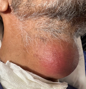
Figure
1:
Anterior cervical mass measuring approximately 15 cm in diameter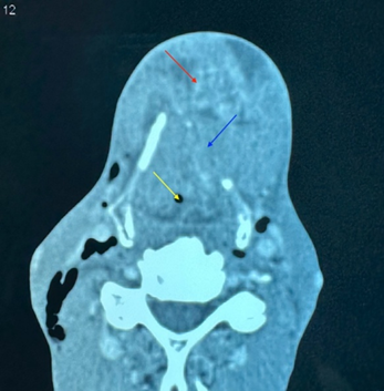
Figure 2: Axial CT scan at the
level of the thyroid cartilage showing a laryngeal mass narrowing the airway
lumen (yellow arrow), with thyroid cartilage lysis (blue arrow) and invasion of
the anterior cervical soft tissues (red arrow)