Cashew sign (cashew nut), a finding of High Relevance in the Diagnosis of Cerebral Venous Thrombosis
Abstract
Is a 64-year-old female patient, with a previous
diagnosis of infiltrating ductal carcinoma with the presence of metastatic
activity at different levels (pulmonary, pleural, meningeal, spine, pelvis).,
who begins suffering with headache of great intensity, associated with
neurological deterioration (seizures, dysarthria) several tomographic studies
are performed during the first month of hospitalization. The second study that
was performed showed an image compatible with the sign of cashew (cashew nut),
which represents a radiological sign of high sensitivity in the diagnosis of
intracerebral haemorrhage secondary to thrombosis of the cerebral venous
sinuses.
Keywords: Cashew sign; Thrombosis; Cerebral Sinuses
Introduction
Cerebral
venous sinus thrombosis is a rare condition whose clinical presentation is very
nonspecific in the early stages. The use of imaging studies in the management
of patients with risk factors is a great aid in early diagnosis that improves
the chances of survival. The cerebral venous sinuses most affected by
thrombosis are the superior sagittal sinus (60%), left transverse sinus (45%),
right transverse sinus (40%) and to a lesser extent, the straight sinus, deep
cortical veins and cavernous sinus1.
The initial radiological findings of cerebral venous sinus thrombosis are
nonspecific, the most frequent is the visualization of homogeneous hyper
densities in the affected sinuses, as well as the “rope sign,” which is the
endoluminal thrombus in the cortical veins2.
In later stages, the cashew nut sign is a highly sensitive diagnostic indicator
of intracerebral haemorrhage secondary to cerebral venous sinus thrombosis3.
Clinical Case
Is a 64-year-old female, diagnosed with
invasive ductal carcinoma of the left breast (November 2020) and presence of
metastatic activity in the lungs, bilateral pleura, spinal canal meninges,
bones (spine, pelvis and right femur) and peritoneum on the left side,
documented in a CT scan of December 2020.
In July 2024, she was taken to the emergency
department due to new-onset seizures. A simple and contrast-enhanced CT scan of
the skull was performed, which showed no evidence of ischemic or haemorrhagic
cerebrovascular events or signs of fracture. No filling defects in venous or
arterial structures were identified at that time. only blurring of the sulci at
the right fronto-parietal level was visualized, with the other findings related
to the aging process (decrease in cortical volume predominantly in the frontal
lobe).
Three days later appears in new CT scan two
hypodense areas at the bilateral frontoparietal level, the one on the left side
with a hyperdense image inside an attenuation range of 40-62 HU, which
constitutes the cashew nut sign, corresponding to a concave intraparenchymal
haemorrhage secondary to thrombosis of the left transverse venous sinus.
Image of a cashew nut or Indian nut (Figure
1), (Figure 2) from a single-phase cranial tomography scan.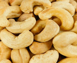
Figure
1: Image of a cashew nut or Indian nut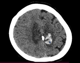
Figure
2: Single-phase cranial tomography scan
Sagittal
(Figure 3) and coronal (Figure 4) images of a single-phase
cranial tomography, showing the cashew sign (cashew nut).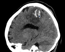
Figure
3: Sagittal image of a single-phase cranial tomography, showing the cashew
sign (cashew nut)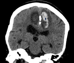
Figure
4: Coronal image of a single-phase cranial tomography, showing the cashew
sign (cashew nut)
Discussion
Cerebral venous sinus thrombosis is a rare type
of ischemic stroke (less than 5% of cases), but with a high mortality rate1.
Risk factors for this condition include
autoimmune vascular diseases, prolonged use of oral contraceptives, severe head
trauma, blood dyscrasias (leukaemia, thrombocytopenia), coagulation disorders
(antithrombin and protein C and S deficiency), as well as oncological
conditions, as presented in this case2.
The pathophysiological mechanism of thrombosis
at this level is related to the hypercoagulable state of the blood secondary to
neoplastic activity and at a distance.
Symptoms are usually nonspecific and related to
the type of vessel affected, the severity and the time of evolution. Severe
headache is the most common symptom (90%), followed by de novo seizures,
paresis, papilledema, signs related to increased intracranial pressure due to
altered cerebrospinal fluid flow and reabsorption (nausea and vomiting) and
altered consciousness3.
The first imaging study that should be
performed is computed axial tomography. It shows changes nonspecific, for
example hyper density of the thrombosed sinuses, hypodensities in the cerebral
parenchyma1.
Summary/Conclusion
The clinical manifestations associated with
cerebral venous sinus thrombosis are nonspecific and often depend on the
patient's medical history to generate a suspected diagnosis. For this reason,
tomography is a cornerstone of diagnosis and treatment, it provides classic
early radiological signs such as hyperdensity of thrombosed sinuses and
hypodensity in the brain parenchyma, which characterize this vascular pathology1.
However, the cashew nut sign is important due
to its high diagnostic (98%) and prognostic sensitivity, failure to identify it
in the early stages delays timely therapeutic intervention and increases the
rate of complications and probability of death3,4.
References
2. Sharma R. Cashew nut sign. En Radiopaedia.org 2022.
4. Ehtisham
A, Stern BJ. Cerebral venous thrombosis. The Neurologist 2006;12(1):32-38.