Clinical Images- Metastatic Anaplastic Thyroid Cancer
65-year-old
female presents to the hospital with shortness of breath that was persistent on
neck and decreased breath sounds in left upper lobe and absent breath sounds on
the right lower lobe as well as tachycardic and hypoxia which improved with 4L nasal cannula oxygen.
Patient underwent CT scan showing 19 mm x 6mm right supraclavicular lymph node (Figure 1) Large soft tissue mass in
the left upper lobe 10.5 x 8.2 x 11.3 cm
with
innumerable diffuse bilateral pulmonary metastatic appearing nodules with areas
of nodular conglomeration (Figures 2 and
3). Patient underwent biopsy revealing metastatic poorly differentiated
thyroid cancer. Poorly differentiated thyroid carcinoma are aggressive tumors
that prompt diagnosis is needed. Samples were sent for next generation
sequencing and patient was planned to start Tyrosine Kinase Inhibitor therapy
as outpatient.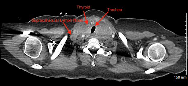
Figure 1: 19 mm x 6mm
Right Enlarged Supraclavicular Lymph Node. Figure also shows positioning of
Trachea and Enlarged Thyroid.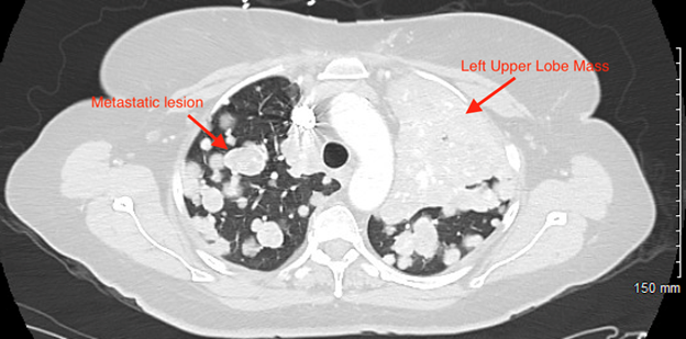
Figure 2: Areas of
Pulmonary Metastasis with a Large Mass in Upper Lung Lobe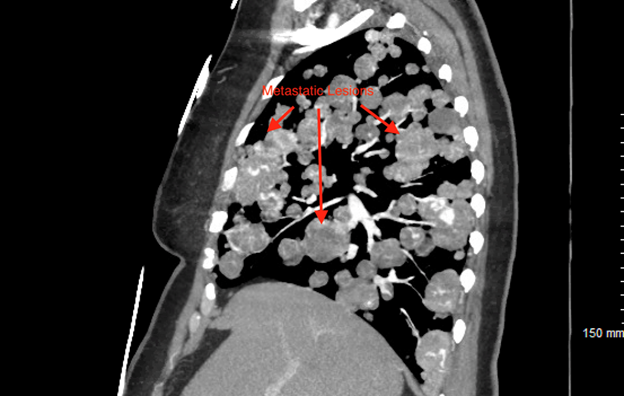
Figure 3: Diffuse
Bilateral Pulmonary Metastatic Appearing Nodules with areas of Nodular
Conglomeration