Gingival Giant Cell Fibroma: Case Report
Abstract
Giant cell fibroma (GCF) has rare occurrences and it manifests as a form of
a fibrous tumor affecting the oral mucosa. Clinically it is presented as a
painless, sessile or pedunculated growth which is usually confused with other
fibrous lesions like irritation. It is distinguished by its unique
histopathological features. Due to the lack of data and rarity of similar
cases, we present a case where a seventy-five-years-old male patient reported
with a nodular growth in relation to the lower mandibular gum. Considering the
size and location of the lesion, excisional biopsy was performed and sent for
histopathological analysis which confirmed the lesion as giant cell fibroma.
Keywords: Giant cell fibroma;
Fibrous tumor; Pedunculated growth; Sessile; Lesion
Introduction
Giant cell fibroma is a unique and rare fibromucosal
mass1.
It was First described by Weathers and Callihan in 19742, the designation was due to its nature compromised of stellate fibroblasts with mononucleated or multinucleated giant cells. Generally, it occurs close proximity to the overlying epithelium. Microscopically a giant cell fibroma is an encapsulated mass of loose fibrous connective tissue that contains numerous characteristic large, plump, spindle shaped and stellate fibroblasts, some of which are multinucleated. These cells are easily observed in the peripheral areas of the lesion3. The most accepted hypothesis for origin of GCF is as a response to trauma or to a recurrent chronic inflammation4, characterized by functional changes in fibroblastic cells, while other cells would take over for collagen synthesis4-6.
GCF is a rare occurrence that picks the interest of doctors. In the following chapter we present a case of an abnormally large GCF and provide an analysis of this oral tumor.
Case Report
A
75 years old male patient chronic smoker and alcoholic addict with a medical
history of diabetes type 2 for 10 years receiving insulin shots, high blood
pressure under treatment in addition to a surgical history; the patient had a
prostate operation under general anesthesia without incidents.
The patient was admitted to our ENT department for a lower gingival swelling the onset of symptomatology goes back to 10 years with the appearance of a progressive lower gingival swelling.
The
whole evolving in a context of apyrexia and conservation of the general state the
oral examination, we noticed the presence of regular mass, firm and mobile on
the lower mandibular gum measuring 3 cm (Figure1).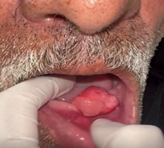
Figure 1: Preoperative image of the patient showing a giant
cell fbroma
The facial CT scan had revealed the presence of an
oval mass well limited with regular borders on the lower gum without any bone
lesions, with multiple calcifications measuring 22* 15 mm extended on 12 mm (Figure 2
Figure 2: Sagital CT scan images showing the tissular mass
with presence of calcifications
An excisional biopsy was performed under local anesthesia and specimen sent for histopathological examination.5 fragments were examined in the biopsy and came out as polypoid. The surface coating was identified as regular sprickle type, without atypia and covered by a slight parakeratosis
The chorion is edematous with a mononuclear inflammatory infiltrate and the presence of giant cells. No signs of malignity were identified.
The histopathological examination had revealed a giant cell fibroma without any signs of malignancies Complete surgical excision of the lesion was performed under local anesthesia and strict aseptic protocol and the specimen was submitted for histopathologic analysis.
Tissue examination under optical microscopy revealed a lesion composed of mature and compact fibrous connective tissue with numerous large spindle- and stellate-shaped mononuclear cells and some multinucleated cells covered by a stratified squamous epithelium with thin, papillary projections.
The stellate-shaped giant cells had hyperchromatic nuclei, while the cytoplasm was well-demarcated and the cells frequently had a dendritic-like process. Areas of inflammation were rarely noted.
No complications or recurrence of the lesion have been noted after 3 months of follow-up.
Discussion
GCF is a rare oral cavity lesion with unique
clinicopathology, which could be diagnosed only on histopathological
examination. Its name is attributed to its histologic presentation, where there
is presence of large multinucleated fibroblasts that tend to occur in close
proximity to the overlying epithelium.
The proliferation and abnormal growth are
likely to be a reaction to the chronic irritation7, inflammation or trauma that induced the functional changes in the
fibrous characteristic of the cells8. Another proposed cause is to be virus
induced, which remains also a possibility7.
Clinically giant cell fibroma is asymptomatic presented as pedunculated or sessile nodule, varying between 0.5 to 1 cm in size. The surface often appears to be papillary and ulcerated. In about 60% of cases, the lesion is diagnosed during the first 3 decades of life and has slight female predilection. It is found more frequently on gingiva followed by tongue and buccal mucosa. Mandibular gingiva is affected twice as often as the maxillary gingiva9.
According to previous studies the GCF doesn’t appear at a specific age however the mean age reported in the studies is late 30s (Table 1). This difference may be linked to its asymptomatic nature or delayed reporting or due to genetic and racial differences.
Also, no significant gender predilection has been determined. It is more frequent on the gingiva, followed by the tongue and the buccal mucosa10.
Histologically GCF is characterized by the presence of numerous large stellate and multinucleated giant cells in a loose dense collagenous fiber which is responsible for the clinical appearance of firm fibroma like mass (Figure 3). The presence of numerous stellate giant cells is what differentiates GCF from other similar lesions11-13.

Figure
3:
Stellate-shaped giant cells with hyperchromatic nuclei and well-demarcated
cytoplasm (H/E original magnification x 200)
Three
main mechanisms responsible for the fusion into giant cells14 are:
· Either the fusion between old and younger cells14
· Or macrophages ingesting same particles resulting in the appearance of multinucleated giant cells14.
The treatment of the CGF consists of surgical excision because the excessive collagenous tissue blocks the tumor regression. Early recognition and complete excision are necessary to minimize repeated surgical intervention.
Conclusion
Giant-cell
fibroma is a rare tumor, distinguished by its peculiar histopathology and its
abnormal growth in the oral and its fibroblastic characteristic that are
responsible for the giant nature of the cells.
Doctors
should be familiar with the different types of fibrous they may encounter
during patient treatment and should note such lesions for further evaluation by
Oral and Maxillofacial Pathologist.
As demonstrated in this case study, GCFs may continue to proliferate until completely removed. GCFs can be treated by curative surgical excision without subsequent recurrence if fully excised.
References
1. Butchi babu K, Nag
S, Hussain MW, Mishra A. Laser excision of giant cell fibroma - A report of a
case and review of literature. Ann Essences Dentistry 2010;2:221-224.
2. Rajendran R,
Sivapathasundharam B. 6th edition. Philadelphia: Elsevier publications. Shafers
Text Book of Oral Pathology 2006:127.
3. Shafer WG, Hine MK
and Levy BM. Shafer's Textbook of Oral Pathology, Elsevier, 6th edition, 2009.
4. Reibel J. Oral
fibrous hyperplasia’s containing stellate and multinucleated cells,
Scandinavian J Dental Research 1982;90(3):217-226.
5. Odell EW, Lock C and
Lombardi TL. Phenotypic characterization of stellate and giant cells in giant
cell fibroma by immunocytochemistry. J Oral Pathology and Medicine
1994;23(6):284-287.
6. Miguel MCC andrade
ESS, Rocha DAP, Freitas RA and Souza LB. Immunohistochemical expression of
vimentin and HHF-35 in giant cell fibroma, fibrous hyperplasia and fibroma of
the oral mucosa. J Applied Oral Science 2003;11(1):77-82.
7. Campos E and Gomez
RS. Immunocytochemical study of giant cell fibroma. Brazilian Dental J
1999;10(2):89-92.
8. Savage NW, Monsour
PA. Oral fibrous hyperplasia’s and the giant cell fibroma. Aust Dent J
1985;30:405.
9. Campos E, Gomez RS.
Immunocytochemical study of giant cell fibroma. Braz Dent J 1999;10:89-92.
10. Reibel J. Oral
fibrous hyperplasias containing stellate and multinucleated cells. Scand J Dent
Res 1982;90:217-226.
11. Nikitakis NG,
Emmanouil D, Maroulakos MP, Angelopoulou MV. Giant cell fibroma in children:
report of two cases and literature review. J Oral Maxillofacial Research
2013;4(1).
12. Okamura K, Ohno J,
Iwahashi T, Netal E. Giant cell fibroma of tongue: A report of case showing
unique S-100 protein and HLA-DR immunolocalization with literature review. Oral
Med Pathol 2009;13:75-79.
13. Santos PP, Nonaka CF,
Pinto LP, de Souza LB. Immunohistochemical expression of mast cell tryptase in
giant cell fibroma and inflammatory fibrous hyperplasia of the oral mucosa.
Arch Oral Biol 2011;56(3):231-237.
14. Williams GT, Williams
WJ. Granulomatous inflammation-a review. J Clin Pathol 1983;36:723-733.