Orbital Cavernoma: Case Report and Literature Review
Abstract
Orbital cavernomas are
the most frequent benign orbital tumor. They are slow-growing lesions of the
intracanal space. A 58-year-old female presented for 7 months of impaired
visual acuity of the left eye associated with severe pain. The physical
examination showed left axial proptosis. Orbital magnetic imaging revealed a 21
mm diameter extraconal space of the left orbital space. The patient underwent
an endoscopic, minimally invasive procedure (FESS) with resection of the tumor
trans nasally.
Keywords: Orbital cavernomas; Orbital tumor; Orbital magnetic
Introduction
The most
common presentation of cavernous hemangioma is a unilateral mass in the lateral
part of the middle third of the orbit1. The radiological work-up
includes Computerized tomography (CT) and magnetic resonance imaging (MRI)2. The surgical
resection is the gold standard and different approaches are possible, including
orbitotomies, transconjunctival access and FESS3,4.
In this
paper, we report a new case of an orbital cavernous hemangioma.
A FESS was performed and after carefully dissecting and resecting the encapsulated tumor, we noted the vascular aspect of the mass with a raspberry color (Figure 1).
Histopathological examination confirmed the diagnosis of cavernous hemangioma.

Figure 1: Preoperative image highlighting the patient's left eye proptosis
Discussion
Estrogen may have a role in the pathogenesis of these tumors, explaining the female predominance with a sex ratio of 7:17. The age at presentation is during the fifth decade of life, with a progressive proptosis associated with diplopia8.
Cavernous hemangiomas demonstrate as well limited, round mass with a hypointense to isointense signal on T1-weighted MRI, while hyperintense on T2 and strongly enhanced after gadolinium injection9. This imaging aspect seems to be an important differentiating aspect of the CSH from other lesions as meningiomas or lymphomas or even other orbital vascular tumors.
The endoscopic approach in treating orbital lesions has been evolving recently and the intraconal tumor surgery is its latest part. The endoscopic management of extraconal tumors is safe and feasible (Figure 2,3), with critical steps such as management of the medial rectus muscle and identifying each orbital structure to avoid postoperative complications10.
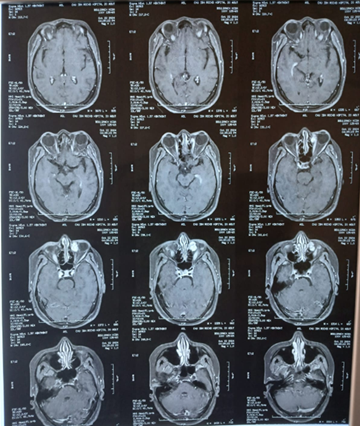
Figure 2: T1-weighted sequence showing progressive enhancement of the left extraconal lesion
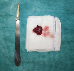
Figure 3: postoperative image of the resected orbital cavernoma
Conclusion
Using FESS to address the orbit has been used for decades in procedures like orbit decompression or dacryocystorhinostomy, but treating orbital tumors such as cavernous hemangioma is a recent field of interest for rhino surgeons.
References
2. Wolin MJ, Holds JB anderson RL, et al. Multiple orbital tumors were cavernous hemangiomas. Ann Ophthalmol 1990;22:426-428.
3. Adrien MT, Ramona G, Torstein MR. Transconjunctival Extirpation of a Voluminous Orbital Cavernoma: 2-Dimensional Operative Video. Operative Neurosurg 2021;20(2):134-135.
4. Rootman DB, Heran MK, Rootman J, White VA, Luemsamran P, Yucel YH. Cavernous venous malformations of the orbit (so-called cavernous haemangioma): A comprehensive evaluation of their clinical, imaging and histologic nature. Br J Ophthalmol 2014;98:880-888.
5. Shah R, Nadimpalli S. Key imaging characteristics for preoperative identification of cavernous sinus hemangioma. Radiology Case Rep 2015;10(1):1013.
6. Linskey ME, Sekhar LN. Cavernous sinus hemangiomas: a series, a review and a hypothesis. Neurosurg 1992;30(1):101-108.
7. Jinhu Y, Jianping D, Xin L, Yuanli Z. Dynamic enhancement features of cavernous sinus cavernous hemangiomas on conventional contrast-enhanced MR imaging. AJNR Am J Neuroradiol 2008;29(3):577-581.
8. Hentati A, Matar N, Dridi H, Bouali S, Jemel H. Bilateral orbital cavernous hemangioma. Asian J Neurosurg 2018;13:1222-1224.
9. Sohn CH, Kim SP, Kim IM, Lee JH, Lee HK. Characteristic MR imaging findings of cavernous hemangiomas in the cavernous sinus. AJNR Am J Neuroradiol 2003;24(6):1148-1151.
10. Lenzi R, Bleier BS, Felisati G, Muscatello L. Purely endoscopic trans-nasal management of orbital intraconal cavernous hemangiomas: a systematic review of the literature. Eur Arch Otorhinolaryngol 2016;273(9):2319-2322.