Sebaceous Cell Carcinoma of the Ocular Region
Introduction
Background: sebaceous cell
carcinoma is a rare and potentially aggressive adnexal neoplasm with
predilection for peri-ocular sites1. Most
commonly, the meibomian gland and the gland of zeis in the eyelids, caruncle
and eyebrow are affected1.
Case presentation: patient
presented with a fleshy growth over the nasal aspect of the left eye of 6
months duration with progressive increase in size and loss of vision in the
eye. Computed tomography (ct) scan done showed a large, irregular mixed density
moderately enhancing left orbital mass. She had a left orbital exenteration
done and histopathologic examination of sections of the tumour showed a
malignant epithelial neoplasm composed of moderate to markedly pleomorphic
cells disposed in nests, cords and sheets invading a desmoplastic stroma and
the sclera. The cells have moderately pleomorphic vesicular nuclei, prominent
nucleoli and foamy to clear cytoplasm with distinct cell membranes (figures 1 and 2). Features are in
keeping with a sebaceous cell carcinoma. She received orbital external beam
radiation to the orbit and systemic chemotherapy and is tumour-free on regular
follow-up post-treatment.
Conclusion: sebaceous
carcinoma is a rare malignant neoplasm, which can clinically and histologically
mimic other benign conditions, thus clinicians and histopathologists require a
high level of suspicion for timely diagnosis. Complete tumor eradication
remains a challenge hence close patient follow-up is critical to identify
recurrence.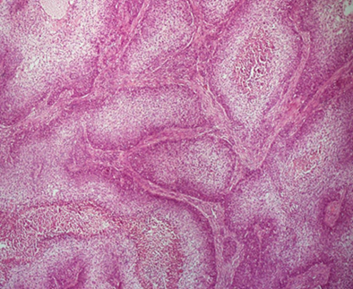
Figure 1. Photomicrograph
showing the histology of sebaceous cell carcinoma: sebaceous epithelial cells
are disposed in lobules interspersed by fibrous septae. Haematoxylin and eosin
stain. X40 magnification. 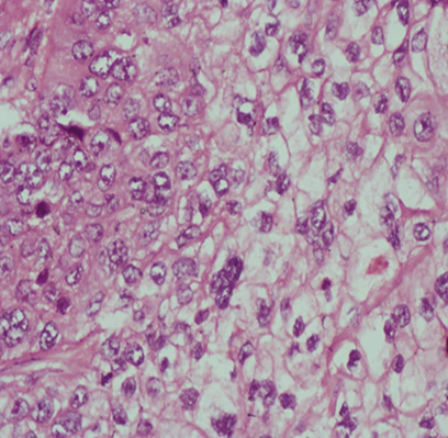
Figure 2. Photomicrograph showing moderately differentiated sebaceous
epithelial cells with abundant foamy to clear cytoplasm. Haematoxylin and eosin
stain. X400 magnification.
References
1. kyllo
rl, brady kl, hurst ea. Sebaceous carcinoma: review of the literature. Dermatol
surg 2015;41(1):1-15.