The Last Hour of Jesus Christ: A Case Study from Recent New Insights on the Turin Shroud
A
paper1 has recently been published
that has presented new insights regarding the blood coming from the Turin
Shroud (TS) and that has posed new interpretations on the possible physical
state of the person who had that blood. In this communication the author offers
a first interpretative cue on the sufferings undergone by Jesus Christ
especially during His last hour on the cross that can subsequently be developed
by any experts in the field interested in the subject. But first let's try to
clarify very briefly some statements that could raise some doubts in the
reader.
The TS is a handmade 3:1 twill linen
cloth, 4.4 m long and 1.1 m wide, on which the front and back images of a human
body are permanently and mysteriously imprinted. According to the current
Catholic Christian tradition, the TS is the burial cloth in which Jesus Christ
was wrapped before being placed in a tomb in Palestine about 2000 years ago and
the author is convinced of this1.
There is evidence that the TS was in
Palestine in the first century A.D., and then taken to Edessa (present-day
Sanliurfa in Turkey). The congruence between the TS face with that of Christ on
Byzantine coins is further evidence that the TS was seen during the Byzantine
empire from the seventh century.
There is evidence that the TS then
appeared in Europe in 1353 in Lirey in France after the Sack of Constantinople
in 1204. In 1988, it was radiocarbon-dated to 1260–1390 A.D2, but the result is questionable and
controversial3-6.
As the process that formed the body image is still unknown, the dating method
cannot be rigorously applied, because the imaging mechanism may, in fact, have
varied the percentage of the TS’s carbon isotopes7. A partial confirmation of this comes from the reduction of nitrogen in
blood samples coming from the TS8.
Regarding the numerous red stains
present on the HS, J. Heller and A. Adler of STuRP (Shroud of Turin Research
Project)9,10 detected the presence of genuine blood from samples
collected from the TS, and this was independently corroborated by the testing
performed by P.L. Baima Bollone11. The body image
reveals many peculiar characteristics that have, thus far, made the body image
unreproducible and not even describable through scientific hypotheses that do
not refer to miracles12,13.
According to the Christian Bible -
and in particular the four Gospels - Jesus Christ, the Son of God made Man, was
severely beaten, scourged, crowned with thorns, had a heavy cross carried to
Mount Calvary where He died crucified. His dead body was then removed from the
cross, wrapped in a shroud and placed in a rock hewn sepulcher where He
remained for about 30-40 hours before rising from the dead. The
author has found compelling scientific correspondences between the narration of
the Bible and the TS which have convinced him that this is the authentic burial
shroud of Jesus Christ14.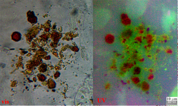
Figure 1: Single
microcytes and small agglomerates coming from sample GF-3EF-3, a piece of
sticky tape put in contact with the bloodstain on the left wrist of Jesus. On
the left, visible epi-illumination on bright field; on the right, the UV
epi-illumination shows the greenish fluorescence of serum surrounding the
microcytes.
1detected on the TS three different types of blood
substances (Figure 1) which, via the
Gospels, can be correlated to different moments of the Passion and Death of
Jesus. The most evident traces appear to be postmortem blood leakage which
probably occurred due to the movement of the corpse during transport and/or in
the tomb and which were transferred into the TS in a fluid phase; less evident
are the pre-mortem bloodstains which probably occurred when Jesus was still
nailed to the cross and which were probably transposed onto the TS after the
blood crusts dissolved due to fibrinolysis in the humid environment of the
sepulcher.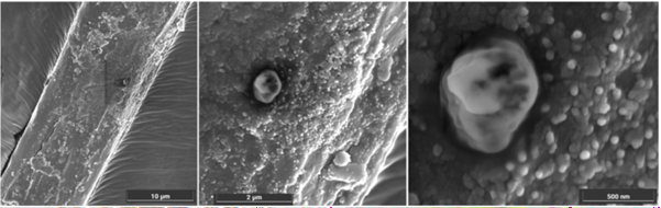
Figure 2: Bloody TS
fiber coming from “h-S” sample: linen fiber coming from Filter-h. Some stacked
microcytes surrounded by creatinine particles can be observed.
Among others, 1reports
the presence of fibrin coupled with erythrocytes and creatinine (Figure 2), which is typical of a
tortured person. But perhaps the most important discovery concerns the fact
that the post-mortem blood of Jesus is composed of microcytes which are
erythrocytes having a diameter of about 0.7 micrometers and which are about ten
times smaller than normal human erythrocytes. Some of these microcytes may also
be residuals of echinocytes.
Experimental results have shown that shrinkage of this type can be
obtained if human blood is diluted with a saturated urea solution. The fact
that Jesus of HS suffered from acute uremia is demonstrated by the atrocious
flagellation, clearly visible on the Relic and clearly mentioned in the Bible,
which would have likely resulted in a renal failure.
But, at this point, it would be
interesting to develop in detail the physical conditions of Jesus on the cross
in His last hour.
The renal (and probably liver)
malfunction or blockage caused microcytic anemia which would have, also, been
exacerbated by a prolonged deprivation of food, imply the extreme difficulties
Jesus had in exchanging oxygen which most likely resulted in extremely labored
breathing. The probable coagulopathy (caused both by the excessive loss of
blood and the coagulation factors) that occurred during scourging, caused
hypovolemic shock. In Jesus’ last hour on the cross before His death,
hypovolemia and severe dehydration (in parallel with John 19:28: “Jesus said,
‘I thirst.’”) would have caused reduced blood flow to His kidneys.
The microcytes would have been
significantly reduced in their ability to exchange oxygen, which would have
resulted in a notable tachycardia that would have been accentuated by tonic and
clonic contractions due to the hypertension of the limbs nailed to the cross.
Jesus’ heart would have been beating very rapidly due to congestive heart
failure while, also, causing a pericardial effusion.
To compensate for these physical
problems in exchanging oxygen, Jesus had to heavily increase His breathing and,
consequently, increase the frequency of His heartbeats, which prompted a heart
attack as His primary cause of death.
In the New Catholic Bible, Psalm
22:15 states "My heart has turned to wax and melts within me." This
seems to provide interesting information about the physical state of Jesus
shortly before his death.
The fact that the "heart has
turned to wax" probably does not only mean that it has taken on the
consistency of wax, but, according to medical pathology, a heart attack is
caused by a significant reduction in blood flow to the infarcted part of the
heart muscle which took on a waxy color too. So, the Psalm confirms what was
experimentally deduced by doctors who studied the TS.
The same Psalm then goes on to say
that the "heart...melts within me," indicating that various causes
"consume" the Body of Christ following renal failure, hemothorax
caused by blunt trauma, and orthostatic collapse15.
Various experts15 confirm that all these causes
produced the early death of Jesus on the cross due to a hemopericardial
infarction. Having established these facts, as mentioned at the beginning, it
would be interesting to take these hypotheses as a starting point to carry out
a more detailed analysis of the particular physical conditions of Jesus Christ
nailed to the cross by comparison with the characteristics of the detected
blood, and to also better understand the extent, still unknown to many, of the
immense suffering that this God-Man declared in the Bible that He would endure
in order to redeem all humanity.
References
1. Fanti
G. New Insights on Blood Evidence from the Turin Shroud Consistent with Jesus
Christ’s Tortures. Arch Hematol Case Rep Rev 2024;9(1):1-15.
2. Damon PE,
Donahue DJ, Gore BH, et al. Radiocarbon dating of the Shroud of Turin. Nature 1989;337:611-615.
3. Rogers
RN. Studies on the Radiocarbon Sample from the Shroud of Turin. Thermochimica Acta 2005;425:189-194.
4. McAvoy
T. On Radiocarbon Dating of the Shroud of Turin. Int J Archaeology 2021;9(2):34-44.
5. Schwalbe
L, Walsh B. On cleaning methods and the raw radiocarbon data from the shroud of
turin. Int J Archaeology 2021;9(1):10-16.
6. Riani
M, Atkinson AC, Fanti G, Crosilla F. Regression analysis with partially
labelled regressors: Carbon dating of the Shroud of Turin. J Statistical
Computing 2012;23:551-561.
7. Phillips
TJ. Shroud irradiated with neutrons? Nature
1989;337.
8. Fanti
G. Could an anomaly in Turin Shroud blood reopen the 1988-radiocarbon-dating
result? World Scientific News 2021;162:102-119.
9. Heller
JH, Adler AD. Blood on the Shroud of Turin. Applied Optics 1980;19(16):2742-2744.
10. Heller
JH, Adler AD. A chemical investigation of the shroud of turin. Can Soci Forensic Sci J 1981;14(3): 81-103.
11. Baima Bollone PL. Indagini identificative su fili della
Sindone. Giornale della Accademia di
Medicina di Torino 1982;1:228-239.
12. Giulio
F. Open issues regarding the Turin Shroud. Scientific Research Essays 2012;7(29):2504-2512.
13. Fanti
G, Hypotheses regarding the formation of the body image on the Turin Shroud. A critical
compendium. J Imaging Sci Technol 2011;55(6):060507.
14. Giulio
F. Why is the Turin Shroud Authentic? Glob J Arch Anthropol 2018;7(2).
15. Bevilacqua
M, Fanti G, D’Arienzo M. New light on the sufferings and the burial of the
turin shroud man. Open J Trauma 2017;1(2):47-53.