Turin Shroud: Insights’ Review Confirming Biblical Reports About Etiology of Jesus Christ’s Death and Resurrection
Abstract
Various new reports have been published on the blood found on the ts (turin shroud). These publications both confirm and improve past hypotheses regarding the particular physical state of jesus during his last twenty hours before dying.
The various accompanying pathologies such as orthostatic collapse, asphyxia, uremia and hemothorax, are accentuated by the insult that produced a high-stress heart disease. Considered individually, the pathologies would have led to death; however, tamponade due to hemopericardium was the primary cause of jesus’ death.
Further, specific analyses of the bloodstains coupled with other characteristics such as rigor mortis and the absence of putrefaction, allowed the author to establish what happened to the body of jesus after being taken from the cross, wrapped in the ts and placed in the sepulcher.
The analysis also revealed some facts that are inexplicable from a scientific point of view and suggested a phenomenon related to the transparency of the matter. The study supports catholic religion belief and therefore, arrives at the hypothesis of the phenomenon of the resurrection to explain what was experimentally detected on the ts. This analysis also provided a physical interpretation of the dogma of faith in the virginity of the mother of god.
Keywords: turn shroud; bloodstains; erythrocytes; fibrin; urea; beta-activity; fluorescence; hemopericardium; rigor mortis; putrefaction; body image; resurrection; dogma
Introduction
Four papers have
been recently published, the first regarding the characteristics of the blood
sampled from the ts1. Consequently,
the physical conditions of jesus christ have been analyzed in the twenty hours
before his death on the cross2,3 and
in the thirty-forty hours after his death4
by merging information coming from the scientific analyses of the ts and
information coming from the chb (christian holy bible). In this review article,
the principal results highlighted in these papers are summarized and commented
on with some improvement.
According to the chb, jesus christ, the son of god made man, was severely beaten, scourged, crowned with thorns and had a heavy cross he carried to mount calvary, where he died crucified. His dead body was then removed from the cross, wrapped in a shroud and placed in a rock-hewn sepulcher where he remained for about 30-40 hours before rising from the dead.
According to pope julius ii who approved the mass and the office of the ts in 1506 and the subsequent catholic christian tradition, the ts is the burial cloth that wrapped the body of jesus christ before he was placed in a tomb in palestine about 2000 years ago, see (figure 1). The catholic christian church does not impose any veneration requirements of the ts, even though science has been unable to refute what is reported by tradition.
The chb synthetically describes what one can observe from a scientific point of view on the ts with more significant details. Still, it also adds that jesus christ was resurrected. The author has found compelling scientific correspondences between the narration of the bible and the ts. Thus he is convinced that this was indeed the authentic burial shroud of jesus christ5.
Refs.4-8, show that the ts is a handmade 3:1 twill
linen cloth, 4.4 m long and 1.1 m wide, on which the front and back images of a
human body are permanently and mysteriously imprinted in a way that has yet to
be reproduced with all its notable characteristics4-7,10-11,
see (figure 2).
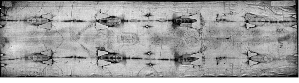
Figure 1: ts
photographed by g. Enrie in 1931.
Fanti’s note: this figure must not be distorced, but
should be put on a double column space, preferably on the top of the page.
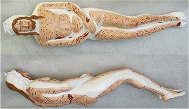
Figure 2:
manikin obtained from a deep study of the ts where the author painted in white
the human body parts not imprinted on the ts due to incomplete wrapping1.
Several features found on the facial image of the ts accurately coincide with those found in depictions of christ on byzantine coins starting from the 7th century a.d. this proves that the ts was seen during the byzantin empire12.
After disappearing during the sack of constantinople in 1204, the “shroud of christ” later appeared in europe in 1353 in lirey in france. In 1532, a fire damaged it at chambéry in france. In 1578, it was taken to turin, where it has remained until now, apart from some short wartime periods when it was hidden.
A piece of linen (officially 7 cm long and 1 cm wide) was cut from the relic in 1988 and given to laboratories in oxford, zurich and tucson (arizona) for radiocarbon dating. Each laboratory measured the 14c/12c isotopic ratio to assign an age to the samples. It was dated to 1260-1390 a.d.13; however the result is questionable and very controversial14-17. The statistical procedure followed and the underestimation of the systematic effects is highly debated17.
As the process that could form the body image on the ts with all its particular features is still unknown, it might quite possibly, be related to a neutron flux18, which potentially varied the percentage of the ts’s carbon isotopes altering what should have been the normal radiocarbon dating result for the linen cloth. In particular, the nitrogen atoms of the ts would have transformed into carbon ones and a partial confirmation of this hypothesis based on a neutron flux comes from blood results of the ts that is poor in nitrogen, see ref.19. In addition, the imaging mechanism may have influenced the percentage of carbon isotopes of the ts, which might have produced an additional non-negligible systematic effect.
The ts shows a particularly unusual double human body image, which still cannot be explained due to its unique combination of features which have, so far, proven to be impossible to reproduce6-11,20-23. Nevertheless, many hypotheses have been formulated22,23 which propose how this image may have been formed. According to the author and other experts, the most plausible explanation is that it was produced by an energy of unknown origin of an electric type20,24, probably connected with the holy fire of jerusalem, which reacted with the linen of the ts. The irregular distribution of radiation along the surface of the ts would have produced both the body image and an isotopic alteration of the atoms in the fabric.
Blood on the
ts
On the ts, there
are hundreds of reddish spots of varying shapes and sizes from centimeters to a
few decimeters, almost completely overlapping the imprinted body image. Ref1 has recognized these spots as blood perfectly
consistent with the different types of torture suffered by jesus, wrapped in it
as a corpse.
The problem of the experimental identification of the different reddish bloodstains present on the ts was initially addressed in 197325 when some reddish specimens were removed from the cloth, sampled and studied by a commission of experts. They were the first in the world to identify blood particles in the samples taken from ts linen threads, although they did not recognize what they were seeing. In fact, the final result was inconclusive about whether or not blood was present on the samples. In this conclusion we read, “we detected [among other] rounded or oval bodies of 0.5 - 0.7 microns in which an external capsule, a membrane and an opaque central portion are evident [and] rounded bodies of 2 microns in diameter apparently surrounded by a membrane and made up of fine granular material unevenly distributed and of different electronic density.… the possibility that these formations are red blood cells cannot be excluded ….” These “rounded or oval bodies” very probably correspond to erythrocytes from different blood types and they, in fact, appeared red if viewed with an optical microscope.
This material is composed of elements (carbon, oxygen, potassium, chlorine, sodium, sulfur, iron and others) that are compatible with blood26,27.
Heller and a. Adler of sturp (shroud of turin research project)28,29, as well as others30, detected the presence of genuine blood in 1980 by testing reddish particles that had been sampled from some sticky tapes put in contact with the ts. They focused their analysis on the chemical compounds forming the reddish material under analysis. Gerard lucotte31 detected the presence of erythrocytes up to 13 micrometers in size, but he also found smaller erythrocytes up to 6.5 micrometers in the analyzed ts samples.
In sharp contrast, analyzing the same samples collected by sturp and studied by heller and adler, w. Mccrone32-38 detected no blood whatsoever on the ts, but instead, he identified very small-sized red pigments (like red ochre and vermillion) which he referred to as “sub-micron particles.” Strangely, a famous microscopist like mccrone did not also consider the particles mentioned above already identified in ref.25 which, according to the author, are also very preponderant27. Adds other information about the blood features on the ts, also discussing their still reddish color, opposite to the common dark brown color of the old blood.
Numerous bloodstains1-4,27-31 scattered throughout the double body image of the ts show evidence that jesus was tortured39: there are blood marks all over the body image consistent with pre-crucifixion flagellation, blood marks on the head consistent with a “crown” of thorns, blood marks on the hand and feet consistent with crucifixion and the blood on the chest that evidences a post-mortem wound typical of a spear. All these tortures received by jesus christ are synthetically described in the chb.
The characteristics and genesis of these bloodstains are not yet fully understood. However, l. Cador40 has recently presented the hypothesis that most of these are blood clots transferred onto the hs. In fact, blood exposed to air coagulates from a liquid state within minutes turning into a fresh, moist clot produced by converting fibrinogen into fibrin that forms a grid where red blood cells are trapped. The clot then retracts, exudes serum and slowly dries out.
Cador supposes that a less frequent type of liquid blood, for example, that coming from the holes in the feet and from the wound in the side, stained the ts linen by direct impregnation. Other bloodstains, for example, those of the hand-wrist wound and some scourge marks correspond to clots that exuded serum in their periphery when drying.
Instead, he supposes that most of the bloodstains on the ts reproduce the image of dry, dissolved-again clots. The corpse wrapped in the ts continued to exude water vapor mixed with other substances such as urea. These clots were moistened by the humid atmosphere of the sepulcher, which dissolved the dry clots devoid of serum again. These dissolved clots formed a soft paste that transferred the bloodstains with clear edges on the fabric onto the ts.
Kelly kearse41, to demonstrate the hypothesis of cador, performed some experiments by depositing blood on a piece of skin, drying it for 3-5 hours and then covering it with a linen cloth. He incubated for 12-15 hours at temperatures and humidity levels similar to those hypothesized for a jerusalem sepulcher and as a result, the bloodstains were imprinted on the linen with well-defined edges that were sharp only under high humidity conditions.
It is also interesting to add what f. Zugibe writes42: “a body can continue to bleed postmortem and this blood can be found, depending upon the circumstances, in either clotted or unclotted form. ... However, in scenarios where the deceased experienced a sudden and/or violent death -such as in cases of severe trauma- the blood’s clotting might begin, however in a matter of 15-30 minutes, the blood becomes fluid again”.
Zugibe also points out that there are no firm rules, but he observes that the fluidity in postmortem blood is connected to the presence of fibrinolysins and its presence in the blood of a corpse is associated with rapid death due to violence. He then adds that it is possible that corpses were bleeding or oozing, sometimes even until the following day, concluding that “with jesus’ having suffered a violent death, this could have caused his postmortem blood to remain fluid long enough to stain his burial shroud when he was placed in it.”
According to the author, however, the fact that a particular condition can produce a specific result does not prove that that result can be obtained only in that particular condition. For example, on a strip of linen cloth immersed in a container with freshly coagulated blood and kept vertical, a separation of a denser and red fluid (corpuscular part) from a lighter part (serum), both characterized by very clear edges appears after a few hours. Even a drop of fresh blood, soaked in a dry linen cloth produces stains with well-defined edges, (figure 3).
Instead, a drop of fresh blood, soaked in a linen cloth imbibed in a liquid mixture of myrrh and urea produces a stain with a clear area in the center as evident in some ts bloodstains, (figure 4).
Regardless of the characteristics of the blood in terms of clots mentioned above, three very different types of blood, type a, b and c have been highlighted in ref.1 and these were classified according to their morphological characteristics.
They do not imply
being from a different person but suggest different moments or conditions of
formation. The author hypothesized the post-mortem type a blood shed on the ts
either still on the cross or in the sepulcher when jesus' body was wrapped in
the ts, type b blood consisting of crusts that coagulated on the skin when
jesus was still alive and type c of uncertain origin due to the penury of
material at disposition. They will help with the following argument on the
physical conditions of jesus christ.
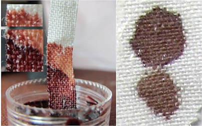
Figure 3: examples of
well-defined bloodstain edges are shown on the left on a strip of linen fabric
immersed in a container of freshly clotted blood and, on the right, on a dry
linen fabric.
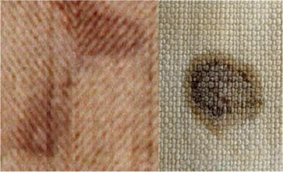
Figure 4: on the left, bloodstains from ts on the forehead. On the right, a fresh bloodstain soaked in a linen cloth imbibed in a liquid mixture of myrrh and urea shows a clear area in the center as evident in the photo of ts on the left.
Blood
evidence consistent with jesus christ’s tortures
Ref.1 added scientific information on the most
important relic of christianity and reported several new features of the blood
there contained, also confirming the atrocious tortures inflicted on jesus
christ that are described in the chb and commented on the refs.6,7,19,25-31.
Among other things, the new features allowed describing in more detail both the actual physical conditions of jesus in the hours preceding his death on the cross and those of the corpse in the subsequent hours until his disappearance, still scientifically inexplicable in the physical details and which will be discussed in the following sections.
Three different types of blood have been detected. This section summarizes the most important novelties1 at the macroscopic and microscopic level detected.
Study of the chest wound: the analysis of the chest wound blood leakage in three different directions allowed confirming the hypothesis that the human body of jesus was turned in different positions, probably during the operations of the corpse on the preparation stone in the sepulcher of jerusalem.
The distinction between the leakage of a reddish part of the blood and an almost transparent part but different from the color of the linen of the ts also allowed developing different hypotheses on the nature of this transparent fluid. According to a spanish scholar, a.s. hermosilla43, it could be a fluid connected to pulmonary edema, but the possibility that this fluid can also be either pleural fluid or serum consequent to hemopericardium must not be disregarded.
Study of the hand-wrist and arm wounds: the analysis of the blood leakage in the hand-wrist area, based on both the color photographs in visible light and ultraviolet, allowed associating the different blood colors with the various origins and characteristics consistent with the result of a square nail section, typical of the romans.
The
pre-mortal blood leakage, which probably occurred on the cross, was
distinguished from the post-mortal one, likely occurred both during either the
transportation or the burial operations and during the dozens of hours of
wrapping the corpse in the linen of the ts. The blood serum leakage was also
evidenced (figure 5).
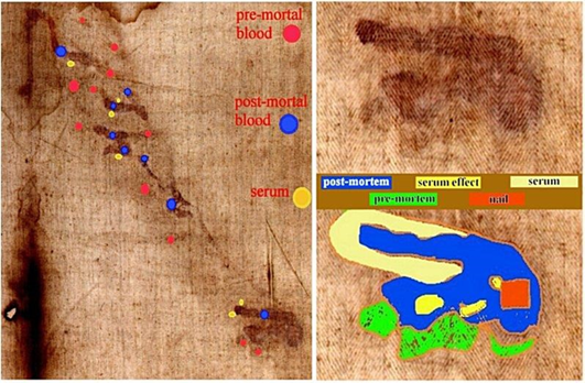
Figure 5: on the left, is a proposed classification of bloodstain types on the left arm of the body image; on the right, is a detailed characterization of different bloodstains on the hand-wrist area1
Bloodstains out of human body image: some blood areas have been identified, almost imperceptible at first glance, but very significant for better understanding the hypothesis of the body image formation.
It is easy to think that the bloodstains were transferred onto the ts linen by contact with the human body, which subsequently transferred the body image. In fact, more than 99% of the ts blood areas are associated with the corresponding body image. However, there are some small bloody areas that a careful eye detects outside the body image, thus posing the question of how they could have formed.
Among these we find the bloodstain outside the body image in the area of the right elbow (frontal image), which clearly derives from a copious outflow of blood from the arm that propagated by flowing along the ts linen.
There are also bloodstains related to whiplash such as the one on the right shoulder highlighted in ref.1 or those corresponding to the knees in the dorsal image which, even if they presuppose contact between the human body and the sheet, do not evidence the corresponding body image. These bloodstains nevertheless are in perfect agreement with the image formation model hypothesized in ref.20 in reference to the effects of an electric field acting on a floating human body, whose image intensity depends on the cosine of the angle between the direction of the electric field and that of the body surface. Therefore in correpospondence of some angles near zero the image approaches zero intensity.
Blood types a, b and c: type a blood, the most interesting for the analysis of the status of jesus christ that will follow, consists of numerous reddish particles present in the adhesive tapes analyzed, see (figure 6), which are reminiscent of erythrocytes or parts of them, present in human blood (7 micrometers in size), but their size, which varies from 0.3 to 2 micrometers, suggests that they are shrunken erythrocytes.
This type of blood can be identified as post-mortem blood that came into contact with fluids probably rich in oils, aloe and myrrh, as well as urea exuded by the corpse, preventing coagulation.
Incidentally, given that
jesus was suffering from very high uremia due to the flagellation which
probably induced kidney (and also liver) failure, this transformation of the
erythrocytes causing microcytic anemia, suggests the extreme difficulties he
had in exchanging oxygen which most likely resulted in extremely labored
breathing.
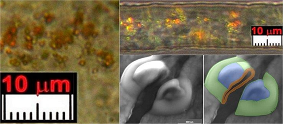
Figure 6: on the left and on the top right, sub-micrometric particles on a linen fiber, with a rounded donut shape from sticky tape put on the feet of the ts dorsal area. On the bottom right, a sectioned sub-micro-metric blood particle from dusts collected at the gluteal area shows a geometry very similar to that of an erythrocyte but with a diameter of about 500 nm1.
Type b blood consists of compact but brittle sherds of crusts darker than type a blood. It is rarer than type a blood and less easily characterized. The few particles found have sizes up to a tenth of a millimeter and have sharp, not rounded edges that suggest previous fragmentations of larger particles, see (figure 7).
This type b blood does not appear to contain the typical microstructures of the red blood cells found in type a blood and one can think that it is pre-mortem blood clotted and dried on the skin from open wounds when jesus was still alive.
Type c material consists of very rare donut-shaped particles found only from the dust of the ts vacuumed from the back side of the face. However, because of a shortage of material, the composition of these particles has not yet been determined.
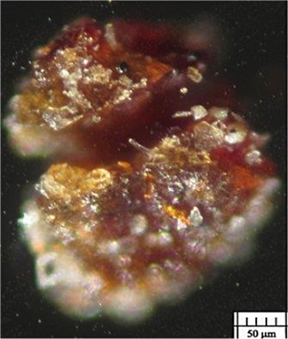
Figure 7: fragmented type b blood sherd coming from the gluteal area1.
Other particles in type a blood: the fact that fibrin particles were found to be arranged along the linen fibers, together with the erythrocytes, confirms that type b blood is indeed blood.
Numerous creatinine particles ranging from 50 to 200 nm were also found arranged in agglomerates that confirm that the blood analyzed is that of a severely tortured person.
Finally, many particles of earthy material were found, typical of that found in jerusalem such as clay and limestone, which support the hypothesis that earthy dust remained on the body of jesus christ because he was not completely washed, but only cleansed.
Fluorescence of type a blood: unlike type b blood, which does not show any kind of detectable fluorescence, type a blood, in addition to a reddish color that is not typical of ancient blood crusts, also indicates a red-orange fluorescence at about 610 nm, which the presence of bilirubin can explain, see the blood sherd of (figure 8). It can be produced by the rupture of erythrocytes during heavy flagellation, with the consequent release of hemoglobin which would then be transformed through the action of liver enzymes into bilirubin.
Even alkalosis produced by an excessive content of urea in the blood due to renal failure2 with consequent hypokalemia or potassium deficiency, can also be produced by muscle spasms, hemolysis and the echinocyte formation induced by pulses in intense electric field, supposed for the body image formation24, should not be forgotten, both in reference to the possible hypothesis of echinocytes formation and the production of free hemoglobin that is then transformed into fluorescent bilirubin.
A new alternative hypothesis could refer to the non-negligible beta-activity of this kind of blood and discussed in the next section, which could have also altered the c14/1988 results.
To confirm that type a blood
is indeed blood, the micro-particles recognized as shrunken erythrocytes are
frequently drowned in sherds of serum showing a greenish fluorescence.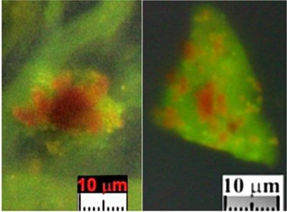
Figure 8: on the left, orange-red
fluorescence of a blood sherd over linen fibers showing a greenish fluorescence
coming from the feet of the dorsal area of the ts; on the right, orange-red
fluorescence of blood drowned in serum showing a greenish fluorescence.
Beta radioactivity of type a blood: the radioactivity measurements performed on the type a and type b blood samples are still preliminary due to the small amount of material available. Nevertheless, information that the author considers innovative and scientifically very interesting has already emerged, demonstrating that ts is selectively radioactive
In particular, type a blood has shown a beta radioactivity of several orders of magnitude higher than one might expect from a common human blood sample or from that produced by the glass slides containing the analyzed samples; however, it has not yet shown gamma radioactivity.
At the moment, there does not seem to be a scientific explanation for this evident anomaly, also because, in the absurd hypothesis that the entire ts had been placed for centuries near highly radioactive material, uniform radioactivity should result, which has not been found. In fact, type b blood, unlike type a blood, despite having shown gamma radioactivity higher than normal, has not shown any kind of beta radioactivity detectable over the background noise.
It will be extremely interesting to perform radioactivity analyses, in the near future, on non-microscopic samples to better define the phenomenon under examination. However, the fact that intense beta radioactivity is found for the type a blood sample clearly calls into question the result of the 1988 radiocarbon dating of the ts since a radioactivity of the material can alter the natural isotopic percentages of the sample under examination, even making it younger by millennia.
Post-mortem erythrocytes stacking of type a blood: ref.1 reports, "in some cases, … blood particles … are observed as not scattered on the surface of the linen fiber, but stacked together. This fact is in agreement with their beta-activity … which demonstrates that electrically charged erythrocytes tend to arrange themselves along the lines of the electric field hypothesized in the sepulcher of jerusalem.”
It must be added that,
according to ref.44 the “pseudo-coagulation
post-mortem” of the blood also causes phenomena of “erythrocyte stacking” and
of “serum expulsion”, see (figure 9).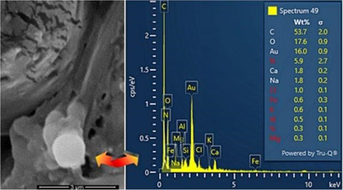
Figure 9. On the left, stacked erythrocytes are indicated by the arrow and, on the right, the elemental analysis shows the similarity to common blood with the effect of the sample metallization with gold1.
Etiology of the death of jesus christ
Type a blood and, in
particular, the microcytes found in1 have allowed the author to
clarify and better develop the various phases of the etiology of the death of
jesus christ.
The pathology that led to the death of jesus was not sudden but most likely developed slowly during the last twenty hours of his life prior to his heart attack that was followed by hemopericardium2,3. This pathology is divided into the phases discussed below.
Last supper: during the last supper [john 13:1-3] it was extremely humiliating for jesus to sit at the table with judas and share his cup with him - knowing that judas would soon betray him. This could have produced intense psychological stress that resulted in the onset of his heart complications.
This syndrome, called stress heart disease (“crepacuore” in italian) ref.45, that could lead to a relatively slow death, is also found at the autopsy table via hematomas of the ventricular surface of the heart with crumbling of the cardiac muscle tissue with visible micro or macro tears in full thickness.
Gethsemane: jesus went then to pray in the garden of gethsemane, where an intense agony caused him to experience hematohidrosis [luke 22:44], a spontaneous emission of blood mixed with sweat from sweat glands due to a rupture of the capillaries surrounding them. It occurs in conditions of intense emotional stress46.
This description from the chb provides evidence that jesus experienced a copious loss of blood that must be taken into consideration in the hypothesis predisposing jesus to infarction
Beatings: according to the chb, several hours passed after judas’ traitorous kiss when jesus was arrested and taken on foot for kilometers to be judged before the sanhedrin, presided over by caiaphas, high priest of the temple of jerusalem. Jesus was then taken to pilate, herod and then again to pilate.
Jesus was severely beaten
during these hours [mark 15:19, isaiah 50:6]. Observed on the ts, as
confirmation of these descriptions, the swollen right cheekbone, the broken
nose and the right beard torn, see (figure 10).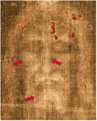
Figure 10. Negative face of jesus on the ts on which the bloodstains have been superimposed in positive by the author. The arrows indicate the broken nose, the swelling on the right cheek and the torn right beard2.
Scourging: jesus was then severely scourged; the ts shows more than 370 scourge wounds47, but in reality, there were likely many more, perhaps even upward of 600. This is because on the relic, only the parts, where the sheet was in contact with the body, show these bloodstains. Therefore the lateral areas of the legs, arms and chest that were undoubtedly hit by the flagrum, because of their position, could not transfer the scourge signs on the ts by contact. During this atrocious torture, jesus was also struck on the head by the scourge, see ref.1.
This astounding number of
wounds not only resulted in a significant blood loss, but also physically
weakened the body already debilitated by the previous events. At least four of
these scourge wounds can be distinguished directly on jesus’ “cardiac aia” (surface
projection of the heart onto the chest wall) which undoubtedly caused
contusions see (figure 11). Such blows from a flagrum did not lacerate
the cardiac muscle. Still, they could have seriously damaged it as a traumatic
insult, causing the onset of the heart attack.
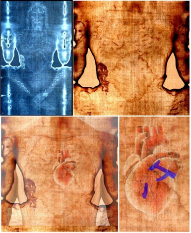
Figure 11: on the top are images of jesus christ on the front side of the ts; on the left bottom, the position of the heart on the ts image and, on the right, evidenced in blue four scourge marks that struck the heart.
Carrying the cross: the immense effort undergone by jesus when he carried the heavy cross to calvary was of no help for the heart partially torn by the congestive heart failure also induced by the violent flagellation. The assumption is made that this stress accentuated the cardiovascular dysfunction and that there was an exacerbation of the heart problems when jesus fell to the ground repeatedly. Perhaps the cross he was carrying bounced off a stone in the ground, causing a dislocation of his right arm. There is confirmation of this in the ts when observing the marks on jesus' shoulders caused by carrying the whole cross and the 3.5 cm dislocation of the right arm in the body image, see ref.48.
Crucifixion on the mount calvary: on the mount calvary, jesus was nailed to the cross by his hands and feet after immense suffering. The holes made on the patibulum (the horizontal beam of the cross) were placed at a greater distance from the limbs, which consequently had to be pulled with ropes, with dislocation, to make their nailing point coincide with the hole in the pre-formed wood39.
The presence of agglomerates of creatinine particles in the type a blood samples confirms the torture endured by jesus. Furthermore, blood composed of microcytes (erythrocytes about 0.7 micrometers in size) is typical of human blood diluted with urea, confirming the acute uremia deriving from the flagellation, which would have likely resulted in sudden renal failure.
The renal and probably liver malfunction or blockage (consequence of renal dysfunction and acute congestive heart failure) caused microcytic anemia that also would have been exacerbated by a prolonged lack of hydration and food deprivation (further support the extreme hypovolemia) that caused difficulties for jesus to exchange oxygen. Therefore, this pathology resulted in labored breathing that further fatigued the heart and accentuated the initial heart injury2.
Effects of tortures : the probable coagulopathy, also caused by the excessive loss of blood that occurred during hematohidrosis and scourging, the hypovolemic shock and the severe dehydration [john 19:28], would have caused reduced blood flow that further burdened the heart.
The microcytes found in ref.1 would have significantly reduced their ability to exchange oxygen, which would have increased the tachycardia significantly, which, therefore, accentuated the heart injury.
The lactic acid produced by the limbs stretched out in the cross would have produced intense tonic and clonic contractions. Jesus’ heart would have been beating very rapidly due to congestive heart failure while also causing a pericardial effusion.
To compensate for these physical problems in exchanging oxygen, jesus had to heavily increase his breathing and, consequently, increasing the frequency of his heartbeats.
All the physical tortures suffered by jesus produced several severe pathologies, each potentially fatal, among which were the hypovolemic shock hypothesized by zugibe and the hemothorax caused by the blows of scourging. It must be considered, in addition, the continuous spiritual agony suffered by jesus accentuated by the innumerable insults received [psalm 69, 20] increased the stress of heart disease. However, the primary cause, according to the authors, is reported in the next section.
The chb [psalm 22:15] states, "my heart has turned to wax and melts within me." this seems to provide interesting information about jesus' physical state shortly before his death. The fact that the "heart has turned to wax" probably does not only mean that it has the consistency of wax but, according to medical pathology, a heart attack is caused by a significant reduction in blood flow to the infarcted part of the heart muscle which took on a waxy color too. The same psalm then says that the "heart ... Melts within me," indicating that various pathologies depleted the body of christ.
This description, which should be confirmed and explored in the near future, allows us to better understand the extent, still little known to many, of the immense suffering that this god-man, as declared in the chb, had to endure to redeem all humanity.
Heart attack with hemopericardium: after all this physical and spiritual torture, it is not difficult to think that a heart attack could have occurred even in a robust person such as jesus christ must have been.
Due to cardiac insufficiency, thrombi could have originated in the so-called "auricle" or atrium, annexed as an offshoot of the left atrium, a small sac-shaped formation, embryonic residue, as highlighted49.
The very high frequency of heartbeats produced a heart attack which caused a significant effusion of blood into the pericardial layer producing hemopericardium with consequent cardiac congestive tamponade and immediate death after a sharp pain in the chest2. This is also in agreement with the chb, which reports the strong cry of jesus shortly before dying [mark 15:37].
Serum and red portion of erythrocytes with fibrin could just be the “blood and water” [john 19:34] that came out of the wound in jesus’ side when the roman centurion struck it to confirm his death.
Events that followed the death of jesus christ
After the
description of the causes of death of jesus christ, an attempt is made here to
describe the various events that followed death by heart attack and ensuing
hemopericardium4.
Rigor mortis of the
body of jesus : the ts shows that the corpse of jesus,
when buried, was in rigor mortis at the time of body image formation50, 51, but this rigor is
not typical of a supine man. His head bowed forward, his knees partially bent.
His feet stretched forward at an angle greater than any movement permitted by
the ankle (and therefore dislocated), following the insertion of the nail, are consistent
with the vertical position of a crucified man, see (figure 12). His
arms, with the hands crossed on the pubic area, were evidently moved after
death to allow entry into the narrow door of the tomb; it appears that a
dislocation weakened the assumed cadaveric rigidity.
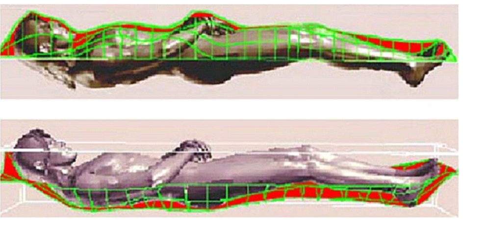
Figure 12: superposition of a numerical model of the ts with a moving digital manikin to determine the best position for wrapping jesus on the ts linen fabric4.
More than a dozen years ago50 the author verified the pronounced cadaveric stiffness of jesus by superimposing a numerical copy of the double imprint of the ts body image with an anthropomorphic numerical manikin (figure 12).
Recently t. Pappas52 confirmed such rigidity, stating that no more than one hour after death, the body of jesus, still hanging on the cross, reached its peak of rigor becoming almost rock-hard in that particular position. This fixed state of rigidity progressively diminishes until it disappears completely due to the action of proteolytic enzymes and the body’s overall process of cellular breakdown, which corresponds to the cadaver’s putrefaction. Rigor mortis can normally last 36-48 hours postmortem, but in some cases, it can reach up to 120 hours in the lower limbs53.
The chb confirms that, after death, the corpse was removed from the cross, wrapped in a shroud and placed in a rock-hewn sepulcher from friday evening to sunday morning. This time period, 36-40 hours [john 19], is coherent with medical reports.
Transport of jesus' body to the sepulcher and wrapping in the ts: it is easy to think that the body of jesus was taken down from the cross and then placed on a first shroud used for transport to the sepulcher; it was then placed on the preparation stone covered with powdered anti-putrid substances, such as aloe and myrrh and half of the ts on top of them4.
According to jewish law, the body of jesus was probably not cleaned of post-mortem blood, but only cleansed of pre-mortem blood, such as that produced by scourging. The rivulets corresponding to the arms and feet due to the unnailing procedure and the leakage of blood and serum from the chest wound related to a spear thrust, are classified as type a post-mortem blood left on the ts1.
Jesus christ's body was then most likely covered with an oily mixture of aloe and myrrh (manteca in italian) [john 19:39]. The absence of traces of the body image or of blood drippings on the sides of jesus' body on the ts suggests that rolls of linen bandages impregnated with anti-putrid substances were placed at its sides to preserve the corpse from putrefaction better.
Absence of movements after deposition in the
sepulcher: after jesus was placed
on the sepulcher's preparation stone, there was no additional relative movement
between the body and the ts4. In
fact, no bloodstain on the ts has any smearing (figure 13 left) because
the bloodstains are perfectly transferred onto the linen fabric, see ref.54 and the type a blood of ref.1 soaked into the ts, was at least partly in a
liquid state (figure 13 right). In other words, once the ts was put in
contact with the body, there was no further relative movement and, therefore,
the body remained in that position on the preparation stone.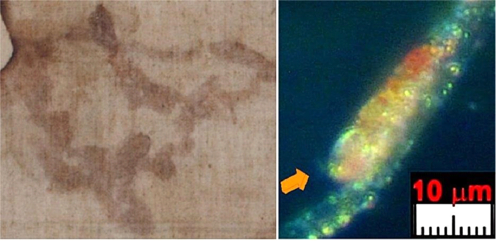
Figure 13: on the left, “belt” bloodstain on the back is an example of bloodstains perfectly transferred onto the linen fabric without minimal smearing. On the right, bloodstained fiber from the wrist sticky tape 3ef. The arrow indicates the drop of blood deposited on the linen fiber in a liquid state4.
This observation leads us to understand that with the evening of good friday approaching and knowing that during the sabbath, the jewish religion forbade any burial operation, those in charge of christ’s burial left the corpse wrapped in the ts on the preparation stone, waiting for sunday to place it in the burial niche of the sepulcher.
No putrefaction signs for the body of jesus: the double body image of the ts clearly shows the absence of signs of putrefaction. In fact, the beginning of putrefaction in a human body occurs with the release of gases, through the orifices, such as methane ch4, carbon dioxide co2, phosphane (or phosphine) ph3 and hydrogen sulfide h2s55, the last two are very aggressive and should have stained the ts. For example, the face, in correspondence with orifices such as the mouth and nose, shows the complete absence of stains4.
Consequently, we can deduce that the body of
jesus remained wrapped in the ts for a time shorter than the release times of
putrefactive gases (bloated stage), estimated between 48 and 72 hours at room
temperature56.
From the chb, findings show two important
confirmations:
·
Jesus christ
died on good friday at about 3 pm and resurrected on easter sunday morning; the
36-40-hour interval corresponds precisely to the time interval highlighted for
the body of jesus to remain wrapped in the ts.
· We read: “because you will not abandon me to the realm of the dead, nor will you let your faithful one see decay.” [psalm 16:10] confirming the absence of putrefaction for the body of jesus wrapped into the ts.
Jesus' exit from
the ts : it results in what follows from the previous sections.
·
The corpse
of jesus was in rigor mortis at the time of the formation of the body image, no
later than 38-48 hours after death.
·
Jesus
remained wrapped in the ts for no more than 36-40 hours because the double body
image of the ts lacks signs of putrefaction.
· The lack of bloodstain smearing shows the complete immobility of the corpse wrapped in the ts therefore the body was not tampered with or moved. Some minimal sliding of the blood clots on the fabric should be detected, especially in the area of the back and glutei if the corpse was moved
By combining this evidence, one must arrive at a deduction that is, for the moment, scientifically absurd: the body of jesus passed through the ts without compromising it materially4. In other words, the physical body of jesus became transparent compared to the linen fabric of the ts!
J. Jackson arrived at this deduction in 199057 when he proposed the hypothesis of a collapsing ts due to gravity force, into a materially transparent human body
As in the present case, the deduction goes beyond current scientific knowledge. Research could stop here but instead, it encourages opening new scientific horizons.
The author, also driven by the perfect congruence determined between what is read in the chb and what is found in studying the ts, tries to find some explanation of what may have happened in the sepulcher when jesus came out of it. The following section will discuss this hypothesis4, for the moment out of science, that agrees with what is reported in the chb where the resurrection of jesus christ is widely mentioned.
Resurrection of jesus christ: the ts does not provide much evidence regarding jesus' exit from the sepulcher. Still, one can nevertheless find some indication in the previous section that the body of jesus passed through the ts without compromising it materially.
In addition, the double body
image then, which is not yet fully scientifically explainable, finds its best
explanation in the presence of an intense energy source, even of the neutron
type19, that occurred during a phenomenon that is still
scientifically inexplicable. Still, one can easily correlate to the
resurrection from the dead. As already seen, j. Jackson57 had predicted all of this, albeit with slightly different
hypothesis.
Some passages of the chb describing facts related to jesus christ’s resurrection and how he manifested himself still alive after his death on the cross follow.
· When the apostle john [20:8-9] entered the tomb on easter sunday, "he saw and believed. They still did not understand from scripture that jesus had to rise from the dead." this very synthetic sentence indicates that the apostle saw the tomb empty upon entering it, but most likely, the arrangement of the ts on the preparation stone made him the first to believe in jesus’ resurrection (figure 14).

Figure 14: above, the ts wrapping jesus' body on good friday. Below, the apostle john, who was present at the burial on good friday when he entered the sepulcher on easter sunday, saw the ts collapsed but not tampered with and the sudarium still in place as though wrapped around the head became materially transparent4.
·
John [20:6-7] reports that “[they] … went straight into the
tomb. He saw … the cloth that had been wrapped around jesus’ head. The cloth
was still lying in its place, separate from the linen.” John's observation
focuses on the arrangement of the sudarium, which suggests that it appeared to
be as if it was left still wrapped around jesus' disappeared head. At the same
time, the ts was collapsed on the stone58. The apostle emphasizes that the sudarium, probably
stiffened by the aromatic substances such as aloe and myrrh, still remained
wrapped around the disappeared head of jesus who had already become materially
transparent and had already caused the collapse of the ts4.
·
The apostle luke (24:30-31) relates that jesus appeared after
his death first to two disciples from emmaus, who ran to warn the apostles.
·
On easter sunday and the following sunday, jesus appeared
twice to the apostles in the closed-door cenacle and, to prove that he was not
a ghost, he dined with them [john 20:19, 20:26].
Hypothesis out of science: transparency of the matter: from what has emerged above, one must consider a hypothesis that goes beyond science, namely that the material body of christ passed through the ts in a "transparent" way and, according to the chb, subsequently passed through the walls of the cenacle.
To explain the phenomenon of transparency with respect to matter, one can think that the diameter of atoms is approximately 10,000 times larger than their nucleus and that, therefore, each atom is practically composed of empty space (if we exclude the very small electrons)4, just as the space of our solar system is practically entirely empty, occupied only by the sun and planets.
Let's consider, for example, a finger resting on the surface of a table. Both are made of atoms, but the finger cannot penetrate the table because the forces that bind the nuclei to the electrons of our finger and the table are much greater than the pressure exerted by the finger on the table itself.
Now, suppose supplying the atoms of the finger with extremely intense energy in the form of photons and therefore, light. This supplied energy could increase the kinetic energy of the individual particles to the point that their kinetic forces exceed those of the interatomic forces. In this particular case, one can think that the atoms can pass through each other and one could also verify the interpenetration of the different materials, such as, for example, the finger with the wood of the table.
The impact probability among protons, neutrons and electrons and therefore their probable destruction, during the crossing would be very small given their extremely small volumes compared to the entire atom in question.
Therefore, in these highly energetic conditions, the interpenetration of matter would not be impossible if perhaps an extremely high light-energy were supplied to the system in question.
It can, therefore, be thought that a physical body is not deprived of its material reality even when it reaches this particularly energetic state, but rather that this body becomes transparent with respect to the surrounding matter and that it can consequently penetrate it as long as it is rich in energy, but that it can then return to its initial state if this light-energy fades over time as chb reported for jesus christ [john 20:19, 20:26].
Although not yet scientifically proven, there are some confirmations of this hypothesis. For example, after the mass dedicated to unborn children on june 17, 2007, in the basilica of guadalupe, the image of the madonna depicted on the tilma showed an intense light emanating from her embryo-shaped womb59. Similarly, a pilgrim photographed the statue of the mother of god at the blue cross in medjugorje which showed a diffuse luminescence, but with a notable intensity of this light concentrated in correspondence with the womb, signifying the presence of the nascent son60, see (figure 15).
Another example concerns metallic materials such as iron. If we consider for example an iron nail at room temperature, we see it opaque to light and therefore the photons that hit the surface of the iron do not pass through it. However, if we heat it to a temperature of around 1000 °c and therefore supply it with thermal energy, the same nail changes color, becomes red and we can observe some photons emitted from its innermost parts of it. This phenomenon occurs because by heating the iron, we supply it with additional thermal energy and this high energy allows the internal photons of the nail to pass through the material thus producing that "transparency of matter" that we do not perceive at room temperature
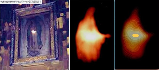
Figure 15: on the left, the mother of god on the tilma of guadalupe with an intense light on her womb. On the center the same intense light from a statue at medjugorje (blue cross) with light contour lines on the right evidencing the light concentration in correspondence of the womb.
Concluding remarks
The recently performed
macroscopic and microscopic analysis of the ts blood1 highlighted characteristics capable of better detailing the
physical conditions of jesus christ, both during the atrocious tortures
suffered before the crucifixion and during the wrapping in the relic until the
moment of the resurrection.
Having determined the stunning correspondence between what results from the scientific analysis of the ts and what is reported in the chb, the paper then plausibly describes from a medical point of view the passion, death and resurrection of jesus christ by merging this information.
The various accompanying pathologies such as orthostatic collapse, asphyxia, uremia and hemothorax, accentuated by the insult that produced a high-stress heart disease, if had been considered individually, would have led to death. However, tamponade due to hemopericardium was the primary cause of death and both beatings and flagellation with at least four scourging marks to the heart increased the broken heart syndrome. The culminating moment of congestive heart disease with the muscle-weakening happened when all these pathologies accentuated the micro-fissures of the muscle and, above all, the microcytic pathology caused the labored breathing and tachycardia that led to the rupture of the heart with consequent hemopericardium and cardiocirculatory arrest. We can, therefore, affirm that jesus physically gave his heart to humanity.
Curiously, the fluorescence of type a blood with its beta-activity atypical for blood can in some way confirm the description of some mystics such as a. K. Emmerick and m. Valtorta of the luminous wounds of the glorious body of jesus4.
This paper then describes the condition of the human body of jesus christ after death by examining the rigor mortis, the transport to the sepulcher and wrapping in the ts, the absence of movements after deposition in the sepulcher, the absence of putrefaction signs, the scientifically unexplainable exit of jesus from the ts and the resurrection.
The rigor mortis, the lack of signs of putrefaction and the perfection of the bloodstain decals on the ts relative to blood still in a liquid state demonstrated that current science, in addition to not being able to explain the double body image formation, is also not able to explain how that human body came out of the ts.
Therefore, an attempt to explain these facts has been made by providing a hypothesis that is still outside of science: the possible transparency of matter when it is supplied with a very large amount of light-energy, also frequently reported in the chb. Suppose we extend this hypothesis to other scientifically inexplicable facts, starting from the one that jesus came out of the ts without disturbing it. In that case, one can also try to similarly explain how he came out of the womb of his mother4, thus explaining the marian dogma of the "virgin before, during and after the birth"61.
It is curious that m. Valtorta, in 1949, when the science on the ts was at its dawn, wrote62 what follows, attributing this to a jesus ‘message. “my shroud, o mary, for those who know how to see, is not only a testimony that i truly died and rose again, but also a testimony of how i was conceived and born, not according to the laws of humanity. It is therefore a confirmation of the truths that my religion teaches: my conception by the work of the holy spirit; the divine motherhood of mary; her perpetual virginity; my passion and death; my glorious resurrection. But this is a confirmation for those who, in the light of god, are given to see”.
Commenting on this point, religion professor sabrina caruso and writer guillaume nocq observed that the holy spirit has always accompanied jesus christ, the alpha and the omega (beginning and end), both from conception (making the male gamete transparent to the body of the mother of god) and from birth (with the child still transparent with respect to the maternal womb), and then both to death (“father, into your hands i commit my spirit.” [luke 23:46]) and to the resurrection (the corpse of jesus was transparent with respect to the ts). In the same way the holy spirit accompanies each of us throughout all our lives. As it happens for each of us, the divine spirit incarnated through mary during the annunciation and remained in jesus until he gave up the spirit by dying on the cross. He then regained the spirit rising again in the sepulcher and therefore this place became similar to the maternal womb, a place of transition between the divine and human world.
In the end, one might ask
what messages the ts, this simple linen cloth that bears a double bloody human
image, wants to leave us? They seem to be multiple and multidisciplinary and
here are some examples.
·
From a medical point of view1-4, the ts confirms and adds much scientific information about
the torture and death of jesus compared to what is reported in the hcb; in
particular, it clarifies and details the type of atrocious tortures suffered by
the man who was wrapped in it and attests to his death, it makes us understand
how much this man voluntarily suffered for men before dying. It also shows that
that man remained wrapped in it for only 30-40 hours and then disappeared in a
scientifically inexplicable way, which the bible explains to us with the
resurrection.
·
From a more general scientific point of view, the analysis of
the body image, which is not reproducible and even explainable22, 23, makes one think that a particular
radiative-energetic phenomenon not known to science occurred.
·
From the selective radioactivity determined in the ts1 and the reduced amount of nitrogen determined in the blood19, one can think that the phenomenon unknown to science was
probably connected to a neutron source; therefore, the radiocarbon result of
1988 is probably not reliable, but perhaps this result will be the first
scientific proof of the resurrection.
·
The ts is a scientifically singular case. There are countless
facts about the ts in favor of its authenticity, none against5. However, it cannot be demonstrated with absolute scientific
certainty that this burial cloth wrapped the body of jesus christ. The
explanation for the author of this singularity is simple: the ts derives from
god and god always wants to propose himself without imposing himself, to leave
the decision to each of us in the end whether to recognize him or not, thus not
affecting the free will that he has been given to us.
·
The study of the ts is a significant case that demonstrates
that science and faith must confront each other and merge their results to
arrive at the truth. In agreement with einstein: "religion without science
is blind. Science without religion is lame"63.
·
From the fusion of information derived by science and
religion (chb), in the case of the ts, it is possible to confirm that christ
rose from the dead.
· Finally, the ts demonstrates the smallness of man and his science in the face of this relic, which consequently attenuates his arrogance when he discovers scientific novelties: "knowledge not accompanied by the fear of god produces the arrogance that makes man consider his own what is a gift"64
Acknowledgments
The author heartily thanks
the late prof. Don lieto massignani, who highly encouraged the writing of this
paper and the holy spirit; he also thanks all the institutions and scholars1 who provided him with material from the ts.
Many thanks to carol gregorek of shroud science group, who provided helpful advice and improved author’s english language skills.
Ethical statements
The author of the christian
catholic religion was able to significantly strengthen his faith after having
carried out scientific studies on the ts. During these studies, also carried
out in collaboration with scholars opposed to authenticity, the author has
always favored maximum objectivity, which has always confirmed what was
suggested to him by faith
Funding
This research was partially
supported by a religious group that requested anonymity and entrusted the
author with the analysis of the so-called "padre pio handkerchief," a
fabric on which two images considered miraculous are imprinted on the front and
back, of the ts-like jesus christ and of saint pio of petralcina respectively.
Following criticism received in a similar case about the possible bias present in the paper due to the origin of the funds, the author clarifies that that research has been concluded for more than a year and that he himself has decided to use part of the funds directly assigned to him to publish this article.
Conflicts of interest
The author declares no
conflict of interest.
References
1. Fanti g. New insights on blood
evidence from the turin shroud consistent with jesus christ’s tortures. Arch
hematol case rep rev 2024;9(1):1-15.
2. Fanti g. Ascolese m. Turin
shroud: etiology of jesus christ’s death for infarction followed by
hemopericardium. Int clinc med case rep jour 2024;3(9):1-12.
3. Fanti g. The last hour of
jesus christ: a case study from recent new insights on the turin shroud. Medi
clin case rep j 2024;2(3):420-422.
4. Fanti g. Shroud of turin: what happened to jesus
christ’s human body after death? J biomed res environ sci
202407;5(10):1278-1287.
5. Fanti g. Why is the turin
shroud authentic? Glob j arch & anthropol 2018;7(2):555707.
6. Jumper ej, adler ad, jackson
jp, pellicori sf, heller jh, druzik jr, a comprehensive examination of the
various stains and images on the shroud of turin, archaeological chemistry iii,
acs advances in chemistry 1984;205;447-476.
7. Schwalbe la, rogers rn.
Physics and chemistry of the shroud of turin, a summary of the 1978
investigation. Analytical chemical acta 1982;135(1):3-49.
8. Jumper eric j. And robert w. Mottern, scientific
investigation of the shroud of turin. Applied optics 1980;19(12):1909-1912.
9. Mottern rw, london rj, morris
ra. Radiographic examination of the shroud of turin - a preliminary
report. Materials evaluation 1980;38(12):39-44.
10. Fanti g. Hypotheses regarding
the formation of the body image on the turin shroud. A critical compendium. J
of imaging sci technol 2011;55(6):1-14.
11. Fanti g, maggiolo r. The
double superficiality of the frontal image of the turin shroud. J. Of optics a:
pure and applied optics 2004;6:491-503.
12. Fanti g. Byzantine coins
influenced by the shroud of christ. Jenny stanford publishing pte. Ltd.
Singapore 2022.
13. Damon pe, donahue dj, gore bh,
et al. Radiocarbon dating of the shroud of turin. Nature 1989;337:611-615.
14. Rogers rn, studies on the
radiocarbon sample from the shroud of turin, thermochimica acta 2005;425:189-194.
15. Thomas mcavoy. On radiocarbon
dating of the shroud of turin. Int j archaeol 2021;9(2):34-44.
16. Schwalbe l, walsh b. On
cleaning methods and the raw radiocarbon data from the shroud of turin. Int j
archaeol 2021;9(1):10-16.
17. Riani m, atkinson ac, fanti g,
crosilla f. Regression analysis with partially labelled regressors: carbon
dating of the shroud of turin. J statistical computing 2012.
18. Phillips tj. Shroud irradiated with
neutrons? Nature 1989;337.
19. Fanti g. Could an anomaly in
turin shroud blood reopen the 1988-radiocarbon-dating result? World
scientific news 2021;162:102-119.
20. Fanti g. Holy fire and body
image of the holy shroud: divine photography hypothesis. World scientific news
2023;176:104-120.
21. Fanti g. Open issues regarding
the turin shroud. Scientific research and essays 2012;7(29):2507.
22. Jackson j, propp k, rebecca
jackson r, koumis a, bertrand j. The shroud: a critical summary of data,
observations and hypotheses. Turin shroud center of colorado, usa.
23. Fanti g. Hypotheses regarding
the formation of the body image on the turin shroud. A critical
compendium j imaging sci 2011.
24. Fanti g. Can corona discharge
explain the body image formation of the turin shroud? J imaging science technol
2010;54(2).
25. Filogamo g. And zina a. La santa sindone,
ricerche e studi … della commissione pellegrino del 1969, magazine:
suppl. Riv.
Diocesana torinese 1976;56.
26. Fanti g, zagotto g. Blood
reinforced by pigments in the reddish stains of the turin shroud. J cultural
heritage 2017;25:113-120.
27. Fanti g. A reexamination of
the pigment-reinforcement hypothesis of the turin shroud’s bloodstains. World
scientific news 2024;9(1):1-15.
28. Heller jh, adler ad. Blood on the shroud of
turin. Applied optics 1980;19(16);2742-2744.
29. Heller j.h., a.d. Adler, a
chemical investigation of the shroud of turin. Canadian society of forensic
science j 1981;14(3):81-103.
30. Baima
bollone pl. Indagini identificative su fili della sindone. Giornale della
accademia di medicina di torino, nº 1982;228-239.
31. Lucotte
gerard, vérités sul le saint suaire, atelier fol’fer, bp 20047, 28260 anet
2010.
32. Mccrone
w. C. And skirius c., light microscopical study of the turin 'shroud,' i. The
microscope 28, 105 (1980)
33. Mccrone
w. C.: light microscopical study of the turin „shroud‟ ii. The microscope 28,
no. 4 (1980) pp. 115- 120
34. Mccrone
w. C.: light microscopical study of the turin „shroud‟ iii. The microscope 29,
no. 1 (1981) pp. 19-39
35. Mccrone
w. C.: the shroud of turin: blood or artist's pigment? Accounts of chemical
research, am. Chemical society, 23 (1990) pp. 77-83
36. Mccrone
w. C., judgement day for the turin shroud. The microscope pub., chicago, usa
(1997).
37. Mccrone
w. C., shroud 1999, the microscope 47, nº 1 (1999) pp. 55-61
38. Mccrone
w. C., the shroud image, the microscope 48, nº 2 (2000) pp. 79-85
39. Bevilacqua m, fanti g,
d’arienzo m. New light on the sufferings and the burial of the turin shroud
man. Open j trauma 2017;1(2):047-053.
40. Cador l. Mode de formation des
taches de sang du linceul, mntv-montre nous ton visage, 2024.
41. Kearse k. A simple, natural
mechanism for the transfer of dry bloodstains onto the shroud of turin. Int j
archaeology 2023;11(12)17-21.
42. Zugibe
f. The crucifixion of jesus: a forensic inquiry. M evans and co, rowman and
littlefield publishing group, lanham usa. Second edition 2005;214.
43. Hermosilla
alfonso sánchez, “the oviedo sudarium and the turin shroud”, 1st international
congress on the oviedo sudarium and the holy shroud in spain – valencia, centro
español de sindonologia (ces), april 28-30, 2012 valencia, spain.
44. Macchiarelli
l, arbarello p, di luca n, feola t, medicina legale, ii ed., minerva, medica,
torino (italy), 2005, https://art.torvergata.it/retrieve/e291c0d4-385c-cddb-e053
3a05fe0aa144/determinazione%20dell%27epoca%20della%20morte.%20segni%20consecutivi.pdf
45. Wirtz petra h, roland vk. Psychological stress,
inflammation and coronary heart disease, curr cardiol rep 2017;19:111.
46. Maglie r, caproni m. A case of blood sweating:
hematohidrosis syndrome 2017;189(42).
47. Fanti g, malfi p. The shroud
of turin, first century after christ!, jenny stanford pub singapore 2020.48. Bevilacqua m, fanti g, d'arienzo m, de caro r. Do we
really need new medical information about the turin shroud? Injury
2014;45(2):460-464.
49. Bussière jp, bonnet d, renard jl, et al. Contribution
of transesophageal echocardiography in the investigation of the atrium in
systemic embolism. Annales de medecine interne 1992;143(1):5-10.
50. Fanti g, basso r, bianchini g.
Turin shroud: compatibility between a digitized body image and a computerized
anthropomorphous manikin. J imaging sci technol 2010;54(5):050503-1/8.
51. Bevilacqua m, concheri g,
concheri s, fanti g, rodella s. Rigor mortis and news obtained by the body’s
scientific reconstruction of the turin shroud man. Peertechz j forensic 2018.
52. Pappas
theodora a. Indicia of reliability: evidence of rigor mortis and cadaveric
spasm from the body image on the shroud of turin. British society for the turin
shroud newsletter, issue no. 99 -summer 2024,editor michael kowalski.
53. Sugatha m, venkata r.
Assessment of time since death using forensic autopsies based on the presence
of rigor mortis- a cross-sectional study. Int j contemporary medical research.
54. Kenneth s, habermas gary r.
Verdict on the shroud, robert hale ltd., london, uk, 1982.
55. Glindemann d, stottmeister u,
bergmann a. Free phosphine from the anaerobic biosphere. Environ sci pollut res
1996;3:17-19.
56. Abdulaziz m. Almulhim; ritesh g. Menezes, evaluation
of postmortem changes, national library of medicine 2023.
57. Jackson jp. Is the image on
the shroud due to a process heretofore unknown to modern science? Shroud
spectrum international 1991;34:3-29.
58. Stalley
l. Divine testimony -hidden references to the holy shroud in the bible, jenny
stanford pub., singapore, 2025.
59. Kine master,
linaluz oficial canal youtube, inscreva se, milagre! Luz misteriosa apareceu no
ventre da virgem de guadalupe.
60. Medjugorje usa, unusual photos unrelated to
medjugorje.
61. Catechism of the catholic
church - latin text copyright. Libreria editrice vaticana. 1993.
62. Valtorta
m, quaderni dal 1945 al 1950, cap. 674, 20 may 1949, centro editoriale
valtortiano s.r.l. 2006;1045-1950.
63. Catholic ireland, religion and
science 2024.
64. Aghiorita
n, di corinto m, filocalia vol i, piero gribaudi editore, tipolitografia
porziuncola perugia, italy 1995;143