Delayed Supraclavicular Hematoma and Thyrocervical Branch Pseudoaneurysm Following Ultrasound-Guided Supraclavicular Brachial Plexus Block: A Case Report
Abstract
Background: Ultrasound-guided
supraclavicular brachial plexus block permits real-time visualization of the
brachial plexus and nearby structures, but it does not eliminate the
possibility of vascular injury. Clinically significant bleeding after this
block is uncommon and delayed arterial pseudoaneurysm is particularly unusual.
Case Presentation: We
report a case of 84-year-old man with end-stage renal disease on hemodialysis,
coronary artery disease, congestive heart failure, atrial fibrillation,
hypertension and thrombocytopenia underwent a right ultrasound-guided
supraclavicular block before planned arteriovenous access creation. His
platelet count was 86 × 10^3/µL.
Forty-five minutes after the block, right supraclavicular swelling was
identified. Initial bedside ultrasonography showed a hematoma without active
bleeding or visible pseudoaneurysm. After six hours of stable observation, he
was discharged. Four hours later, he returned with increased firmness and pain.
His platelet count was 78 × 10^3/µL
and computed tomography angiography demonstrated a 9 × 7 × 7 cm hematoma with a
central contrast blush suspicious for pseudoaneurysm. On day 5 after the block,
angiography identified a distal thyrocervical branch pseudoaneurysm, which was
treated with Onyx and Optoblock coils followed by ultrasound-guided
percutaneous thrombin injection. He was discharged the following day.
Conclusion: A negative initial bedside ultrasound does not exclude evolving arterial injury in a patient with persistent or progressive post-block swelling. In patients with clinically relevant bleeding risk factors, serial examination and timely escalation to computed tomography angiography and catheter angiography should be considered when symptoms evolve.
Keywords:
Supraclavicular brachial plexus block, Pseudoaneurysm, Thyrocervical artery, Hematoma,
Ultrasound guidance, Regional anesthesia, Thrombocytopenia, Case report
1. Introduction
Ultrasound-guided supraclavicular
brachial plexus block is widely used for anesthesia and analgesia during
upper-extremity procedures. Ultrasound allows the operator to identify the
plexus, subclavian artery, pleura and other regional structures in real time
and it may reduce unintended vascular or pleural puncture compared with
landmark-based techniques. However, ultrasound is not a substitute for detailed
anatomic knowledge, careful needle visualization or post-procedure surveillance1-4.
The supraclavicular region contains
multiple arterial structures that may be encountered during needle advancement,
including the subclavian, transverse cervical and dorsal scapular arteries. In
a 2019 evidence review, large studies of supraclavicular block reported
vascular puncture rates of 0 to 0.4% and no hematomas, while the review
identified no published hematoma case reports after supraclavicular block at
that time1.
Rare bleeding complications have nevertheless been described with brachial
plexus techniques, including hemothorax after supraclavicular block and
hematoma after other approaches5,6.
We report a delayed right supraclavicular hematoma associated with a distal thyrocervical branch pseudoaneurysm after an ultrasound-guided supraclavicular block in a patient with end-stage renal disease and thrombocytopenia. The case emphasizes the limitations of an initially negative bedside study and the importance of reassessing evolving symptoms. The report is organized according to CARE case-report guidance7.
2. Case Presentation
2.1. Patient information and risk
profile
Here we present a case of 84-year-old
male patient with end-stage renal disease receiving hemodialysis, coronary
artery disease, congestive heart failure, atrial fibrillation and hypertension
presented for scheduled creation of a right upper-extremity arteriovenous
access for hemodialysis. He had received dialysis the day before the planned
procedure. Pre-procedure laboratory testing showed thrombocytopenia with a
platelet count of 86 × 10^3/µL;
the remaining coagulation studies were reported as within normal limits. He was
not receiving anticoagulation for atrial fibrillation because of a previous
gastrointestinal bleed while taking warfarin.
2.2. Block procedure
A right supraclavicular brachial plexus
block was performed in the preoperative area for surgical anesthesia and
postoperative analgesia. Standard monitors were used. The skin was prepared
with 2% chlorhexidine and a sterile ultrasound probe cover was applied.
Ultrasound identified the subclavian artery superior to the first rib and the
upper, middle and lower trunks of the brachial plexus. No additional pulsatile
vessels were visualized during the procedure. A total of 30 mL of local
anesthetic was injected incrementally, consisting of 10 mL of 1% mepivacaine
and 20 mL of 0.5% ropivacaine, with negative aspiration performed every 5mL.
The patient did not report paresthesia and remained hemodynamically stable.
2.3. Initial swelling and delayed
presentation
Forty-five minutes after completion of
the block, swelling was noted in the right supraclavicular area after the
patient was transferred to the operating room. Bedside ultrasound demonstrated
a right supraclavicular hematoma without active bleeding or a visible
pseudoaneurysm. A pressure dressing was applied and the planned surgery was
cancelled. The patient was observed in the post-anesthesia care unit for six
hours. He remained hemodynamically stable without evidence of hematoma
expansion and was discharged with close follow-up.
Four hours after discharge, he returned
to the emergency department because of new firmness in the right
supraclavicular area and pain when turning his head to the right. He had no
motor or sensory deficit, respiratory distress or abnormal vital signs. His
platelet count was 78 × 10^3/µL.
Computed tomography angiography of the neck demonstrated a large right
supraclavicular hematoma measuring approximately 9 × 7 × 7 cm with a central
area of contrast blush concerning for pseudoaneurysm formation.
2.4. Definitive diagnosis and treatment
The patient was admitted to the vascular surgery service for continued pressure dressing and neurovascular and wound checks every four hours. He underwent dialysis the following day using a pre-existing right internal jugular tunneled dialysis catheter. On the fourth day after the nerve block, the neurosurgery and interventional radiology teams were consulted. On the fifth day after the block, diagnostic cerebral angiography demonstrated a large distal thyrocervical branch pseudoaneurysm (Figures 1 and 2). The pseudoaneurysm was embolized with Onyx and Optoblock coils, followed by ultrasound-guided percutaneous thrombin injection. The patient tolerated the intervention and was discharged the following day show in Table 1. Longer-term follow-up information was not available in the source record.
Table 1: Clinical timeline.
|
Time point |
Clinical event |
|
Day-1 |
Hemodialysis before the planned arteriovenous access
procedure. |
|
Day 0, before surgery |
Right ultrasound-guided supraclavicular block
performed. Platelet count: 86 × 10^3/µL. |
|
Day 0, 45 minutes after block |
Right supraclavicular swelling noted. Bedside
ultrasound showed a hematoma without visible active bleeding or
pseudoaneurysm. Surgery cancelled; pressure dressing and observation
initiated. |
|
Day 0, 6 hours after observation |
Hemodynamically stable without documented expansion;
discharged with close follow-up. |
|
Day 0, 4 hours after discharge |
Returned with increased firmness and pain. Platelet
count: 78 × 10^3/µL. CTA showed a 9 ×
7 × 7 cm hematoma with contrast blush. |
|
Day 1 after readmission |
Dialysis performed through the pre-existing right
internal jugular tunneled catheter. |
|
Day 4 after block |
Neurosurgery and interventional radiology consulted. |
|
Day 5 after block |
Angiography demonstrated distal thyrocervical branch
pseudoaneurysm. Onyx and Optoblock coil embolization and percutaneous
thrombin injection performed. |
|
Day 6 after block |
Discharged after tolerating treatment. |
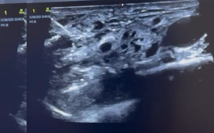
Figure 1:
Computed tomography angiography of the neck demonstrating a large right
supraclavicular hematoma with a central contrast blush concerning for
pseudoaneurysm formation. The airway was patent.
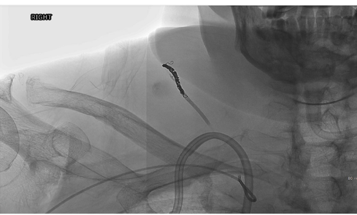 Figure 2: Representative procedural
images from the diagnostic angiography and endovascular treatment of the distal
thyrocervical branch pseudoaneurysm. Panels A-C show the procedural images
obtained during angiography and embolization.
Figure 2: Representative procedural
images from the diagnostic angiography and endovascular treatment of the distal
thyrocervical branch pseudoaneurysm. Panels A-C show the procedural images
obtained during angiography and embolization.3. Discussion
3.1. Bleeding risk and regional anatomy
This case illustrates a rare but
clinically important vascular complication temporally associated with an
ultrasound-guided supraclavicular block. The available evidence suggests that
bleeding after supraclavicular block is uncommon. Tsui and colleagues reported
vascular puncture rates of 0 to 0.4% in large supraclavicular-block studies and
identified no hematomas or published hematoma case reports for this approach in
their review1.
Large clinical series have also reported low rates of clinically significant
complications after ultrasound-guided supraclavicular block8. These data support
the safety of the technique at a population level, but they cannot exclude
rare, high-consequence events in individual patients.
The supraclavicular fossa is
anatomically complex. The subclavian artery is a key landmark, but smaller
branches of the thyrocervical system may not be conspicuous on routine
grayscale imaging. The transverse cervical and other arterial branches can be at
risk during needle advancement1,3.
In addition, anatomic variation in the relationship between the brachial plexus
and subclavian artery has been described and ultrasound may reveal variants
that materially change the safest needle trajectory4. In the present case,
no additional pulsatile vessel was visualized during the block, yet a distal
thyrocervical branch pseudoaneurysm was later demonstrated. This finding
highlights that visualization of the expected major vessels does not prove that
all smaller or variant vessels have been excluded from the needle path.
3.2. Patient-specific considerations
The patient had several potential
bleeding-risk modifiers, including thrombocytopenia and end-stage renal
disease. He was not taking chronic anticoagulation for atrial fibrillation, but
the source record does not report whether anticoagulation was used during the
preceding dialysis session. Therefore, the relative contribution of platelet
dysfunction, thrombocytopenia, uremia, dialysis-related factors and direct
arterial trauma cannot be determined. The case also does not establish that the
needle directly injured the later-identified branch; the relationship is
inferred from the temporal sequence, location of the hematoma and angiographic
diagnosis.
Bleeding-risk assessment before
peripheral nerve and interfascial plane blocks should be individualized rather
than based solely on whether a patient is receiving therapeutic
anticoagulation. Expert consensus emphasizes that evidence for bleeding risk is
limited and that recommendations should support, rather than replace, clinical
judgment1.
In a patient with thrombocytopenia or renal disease, the decision to proceed
should incorporate the urgency and benefit of the block, alternative anesthetic
strategies, the planned needle path, the ability to compress the site and the
feasibility of post-procedure observation.
3.3. Diagnostic implications of delayed
symptoms
The initial bedside ultrasound
identified the hematoma but did not demonstrate active bleeding or a
pseudoaneurysm. Four hours after discharge, the patient returned with new
firmness and pain and Computed Tomography Angiography (CTA) then showed a large
hematoma with a contrast blush. This sequence is clinically important because
pseudoaneurysms may evolve after the initial injury and may be missed when the
study is performed early, when the lesion is small or when the relevant branch
is difficult to visualize. Ultrasound guidance during the block and an
initially reassuring post-procedure examination should therefore not be
interpreted as definitive exclusion of delayed arterial injury.
New or progressive swelling, increasing
firmness, pain, pulsatility, neurologic symptoms, airway symptoms, hemodynamic
change or a fall in hemoglobin should prompt immediate reassessment. When the
clinical course is discordant with an initial bedside study, contrast-enhanced Computed
Tomography Angiography (CTA) can define the size and extent of the hematoma and
identify a contrast blush or pseudoaneurysm. Catheter angiography remains
useful when a treatable arterial source is suspected because it can establish
the diagnosis and permit selective embolization.
3.4. Management considerations
Management should be individualized
according to hemodynamic status, airway and neurologic findings, hematoma
behavior and the anatomy of the arterial lesion. Initial measures may include
close observation, pressure when anatomically appropriate, serial neurovascular
and airway assessment and early involvement of vascular surgery, interventional
radiology and other relevant services. The reported patient was initially
stable, but delayed clinical progression led to Computed Tomography Angiography
(CTA), admission and subsequent angiography.
The distal thyrocervical branch pseudoaneurysm was treated with Onyx and Optoblock coils, followed by percutaneous thrombin injection. Endovascular treatment is commonly used for selected thyrocervical or subclavian branch pseudoaneurysms and branch-vessel hemorrhage, while open or hybrid repair may be appropriate when anatomy, lesion size, collateral circulation or failure of endovascular treatment requires it9-11. In a retrospective series of subclavian and axillary branch hemorrhage, transcatheter arterial embolization achieved 100% technical success and 85.7% clinical success, although the study population and causes of bleeding were heterogeneous11. These results support early consultation with an endovascular team when imaging identifies a potentially treatable branch-vessel source, but they should not be interpreted as a procedure-specific outcome estimate for supraclavicular block complications.
4. Strengths and Limitations
The principal strength of this report is the clinically detailed sequence from block placement to initial ultrasound, delayed return, Computed Tomography Angiography (CTA), angiographic diagnosis and definitive treatment. The case also documents relevant platelet counts and the absence of early neurologic or respiratory compromise. Limitations include the single-patient design, incomplete information about the exact needle gauge and trajectory, absence of documented dialysis anticoagulation details, lack of a long-term follow-up assessment and inability to prove direct needle injury to the pseudoaneurysm branch. The case should therefore generate vigilance and hypotheses rather than establish incidence or causation.
5. Conclusion
A distal thyrocervical branch pseudoaneurysm can present as a delayed enlarging supraclavicular hematoma after an ultrasound-guided supraclavicular brachial plexus block, even when the initial ultrasound does not show active bleeding or a pseudoaneurysm. Thrombocytopenia and end-stage renal disease may increase concern for bleeding, but the specific contribution of each factor cannot be determined from this case. Progressive swelling or new pain warrants serial clinical assessment and timely escalation to CTA and catheter angiography, with early multidisciplinary management when an arterial source is suspected.
6. Declarations
6.1. Ethics approval
This report describes a single-patient
case and was not conducted as human-subjects research. In accordance with
University of South Florida IRB guidance, retrospective review of records for
publication of a single case report involving three or fewer individuals does
not require IRB review or approval.
6.2. Consent for publication
Written informed consent was obtained
from the patient for the collection, analysis and publication of the clinical
information, including images and photographs. Written informed consent was
obtained from the patient for publication of this case report, including all
clinical details and images. The patient was informed about the purpose of the
report, its intended scientific audience and the measures taken to preserve
anonymity.
6.3. Conflict of interest
The authors declare that they have no
financial or non-financial conflicts of interest related to this work.
6.4. Funding
The authors received no external financial support for the work reported in this case report.
7. References
- Tsui
BCH, Kirkham K, Kwofie MK, et al. Practice advisory on the bleeding risks for
peripheral nerve and interfascial plane blockade: evidence review and expert
consensus. Can J Anesth. 2019;66.
- Hanumanthaiah
D, Vaidiyanathan S, Garstka M, et al. Ultrasound guided supraclavicular block.
Med Ultrason. 2013.
- Govender
S, Mohr D, Tshabalala ZN, et al. A review of the anatomy and a step-by-step
visual guide to performing an ultrasound-guided supraclavicular brachial plexus
block. South Afr J Anaesth Analg. 2019.
- Porter
S, Garner HW, Schoch BS, et al. Anomalous course of the brachial plexus
identified during ultrasound-guided brachial plexus nerve block. Turk J
Anaesthesiol Reanim. 2022.
- Singh
SK, Katyal S, Kumar A, Kumar P. Massive hemothorax: a rare complication after
supraclavicular brachial plexus block. Anesth Essays Res. 2014;8(3): 410-412.
- Clendenen
SR, Robards CB, Wang RD, et al. Case report: continuous interscalene block
associated with neck hematoma and postoperative sepsis. Anesth Analg. 2010;110:
1236-1238.
- Riley
DS, Barber MS, Kienle GS, et al. CARE guidelines for case reports: explanation
and elaboration document. J Clin Epidemiol. 2017;89: 218-235.
- Perlas
A, Lobo G, Lo N, et al. Ultrasound-guided supraclavicular block: outcome of 510
consecutive cases. Reg Anesth Pain Med. 2009;34(2): 171-176.
- Neris R, De Camps Martinez ER, Leon E, et al. Unusual
case of an idiopathic thyrocervical trunk pseudoaneurysm: an interesting case
report. Cureus.
2024;16(10): e72273.
- El Naamani K, Abbas R, Sioutas GS, et al. A complication of the transradial approach: thyrocervical trunk pseudoaneurysm with hemothorax. BJR Case Rep. 2023.
- Onishi
Y, Shimizu H, Ando S, et al. Transcatheter arterial embolization of the
subclavian and axillary artery branches for hemorrhage control. Br J Radiol.
2023;96: 20221132.