Holy Shroud in Turin: Bloodstains on the Neck’s Nape and Possible Traces of Myrrh
Abstract
This study investigates the origin of the bloodstains in the occipital region of the Holy Shroud in Turin Shroud (HST), combining image processing and experimental analysis. Through multidisciplinary tests, the formation of these particular blood traces is simulated.
The main findings are the following: it was ruled out that the stains resulted from simple vertical fluid flow; the presence of well-defined angular profile suggests the possible involvement of leaves trapped in the hair after the removal of a crown of thorns made from leafy branches; the light stains with dark borders are consistent with the use of myrrh granules placed between the hair and the cloth; the experimental models, although based on simplified hypotheses, show notable similarities with the original traces of the HST.
Thus, the study proposes new interpretative scenarios and suggests the possibility of future experimental verification using human biological samples, offering a significant contribution to the understanding of this aspect of the Relic.
Introduction
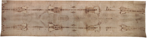
A few studies 1-10, show that the HST is a handmade 3:1 twill linen cloth, 4.4 m long and 1.1 m wide, on which the front and back images of a human body are permanently and mysteriously imprinted 5,6,11,12.
According
to Pope Julius II 13, who
approved the Mass and the Office of the HST in 1506 and declared that it had to
be not only venerated but also adored and the subsequent Catholic Christian
tradition, the HST is the burial cloth in which the body of Jesus Christ was
wrapped before being placed in a tomb in Palestine about 2000 years ago, (Figure
1).
Figure
1:
HST photographed by G. Durante in 2000 (Archdiocese of Turin)
The
Catholic Christian Church does not impose any veneration requirements of the
HST, even though science has been unable to refute what is reported by
tradition.
There are
some indications that the HST was in Palestine in the first century A.D. and
then taken to Edessa 14,15
(present-day Sanliurfa in Turkey) but other scholars hypothesize different
paths for this Relic 16.
Several
features found on the facial image on the HST accurately coincide with those
found on depictions of Christ on Byzantine coins starting from the VII century
A.D. which provides evidence that the HST was seen during the Byzantine Empire 15. After disappearing during the Sack of
Constantinople in 1204, the “Shroud of Christ” then, later, appeared in Europe
in 1353 in Lirey in France. In 1532, a fire damaged it at Chambéry in France.
In 1578, it was taken to Turin where it has remained until now, apart from some
short periods of time when it was hidden during wartime.
In 1988,
radiocarbon dating of the HST yielded an inaccurate date range of 1260-1390 AD 16. This result remains a subject of ongoing
debate 3,17-22, with multiple
studies challenging its reliability due to very probable contamination
especially caused by environmental factors.
Recent
findings, including the detection of Beta radioactivity and fluorescence in the
bloodstains on the HST 1,23-25
further confirm the inaccuracy of the radiocarbon dating results. These
discoveries suggest that neutron reactions related to the body image formation
may have skewed the radiocarbon measurements.
The
presence of selective radioactivity detected in the HST serves as a strong
indication that the 1988 radiocarbon dating results is biased by an intense
neutron flux that altered the isotopic composition of the linen fibers, leading
to a younger apparent radiocarbon age. Such a neutron flux could be easily
associated with the Resurrection of Jesus Christ.
Recent
studies 23,24 have demonstrated
that, from a medical perspective, it is virtually impossible for a medieval
artist to have produced the bloodstains observed in correspondence with the
double-body image on the HST.
These
stains, which exhibit distinct morphological variations, can only be coherently
explained by considering the HST as having been wrapped around a human body
that underwent severe torture and crucifixion in accordance with execution
practices of Roman types, as described in the Christian Holy Bible (CHB).
Moreover,
specific characteristics of these bloodstains, such as the absence of smearing,
further suggest the presence of a phenomenon that remains scientifically
unexplained, potentially pointing to a miraculous occurrence.
Recent
analyses of blood samples collected from the HST 1,3,4,17,23-29
have provided novel insights into the physiological state of Jesus Christ
during His Passion, crucifixion and entombment described in the CHB. One of the
key conclusions emerging from this research is the hypothesis concerning the
mode of Christ’s departure from the HST following the estimated 30 to 40 hours
post-mortem. The study proposes a novel interpretation based on the concept of
material transparency, demonstrating that Jesus' body came out of the HST in a
way that did not touch the integrity of the cloth.
Numerous
studies have been conducted on the bloodstains of the HST 1-11,14,23-29, but curiously, few specific
analyses have been conducted on the characteristics of the bloodstains on the
nape of the neck corresponding to the top of the dorsal image of the HST.
Therefore, this paper analyses these bloodstains through both visible and UV
photos which, among other, present peculiar evidences slightly different from
the others, because they highlight small circular shapes within them that are
not easy to explain at a first sight.
Through
experimental tests aimed to reproduce these peculiar bloodstain marks, this
paper not only tries to give a preliminary explanation to the presence of these
small circular shapes also considering the possible presence of spices
mentioned in the Gospels (Jh 19:39), but also tries to explain some localized
lacks of bloodstains in reference to the possible presence of leaves as hypothesized
by O. Scheuermann 32.
Comments on a Recent
Article About the HST Bloodstains
Very recent
is Ref. 30 which, through
experimental tests, states that in correspondence with the bloodstains “serum
borders would be absent if the body had been washed prior to wrapping it in the
cloth” and concludes that: “These data provide evidence that formation
of serum edges/halos is prohibited under conditions that are characteristic of
post-mortem blood excretion from washed wounds, findings which suggest that the
washing hypothesis is not consistent with what is observed on the Shroud.”
Since the
HST shows both bloodstains surrounded by serum halos and bloodstains lacking
it, it is considered important to better clarify this finding, which is also
useful for the present analysis.
The finding
that the formation of serum halos does not occur for post‑burial bleeding of
blood from washed wounds implies that only some of the bloodstains present on
the HST-specifically, those lacking serum-originate from cleansing of the
corpse (a quick and superficial cleansing because the Sabbath was approaching,
a day of rest for Jews).
In
contrast, those showing serum halos on the HST derive from unwashed bloodstains
likely dripped from the corpse while still hanging on the cross (according to
Jewish law, in fact, post-mortem blood was not to be washed away).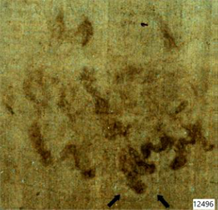
Figure
2:
Bloodstains at the nape of the neck photographed under UV 31. The bloodstain at the bottom centre (see
arrows), shows a halo that could be of serum
Given the
result obtained in Ref. 30, it is
now easier to distinguish on the HST, at least as a first approximation,
between bloodstains with serum-halos attributable to the body still on the
cross (for example that of the chest due to a lance, those of the wrist and on
the feet due to nails and some on the nape of the neck due to a crown of thorns
and some scourge marks on the back) and non-serum bloodstains related to
post-burial bleeding that occurred in the sepulcher after a superficial
cleansing.
It seems
therefore possible to more clearly distinguish which areas were subjected to a
probable quick cleansing and which were not washed in accordance with Jewish
law.
Regarding
the bloodstains on the nape of the neck, (Figure 2) highlights the
brighter outline of a bloodstain that could be traced back to a serum halo.
This would therefore show that this bloodstain on the nape of the neck was not
washed, as one might expect.
Materials and Methods
For the
analyses, the following photos were used: colour photographs taken in visible
light by Gian Durante in 2000 and 2002 and photographs taken by V. Miller in
1978 31, which were recently made
available on the Web (Ref. 31).
As is known, these UV photographs highlight the fluorescence of the serum that
often surrounds bloodstains of the HST and enhance the contrast between the
bloodstains and the background.
To improve
the digital images and thus highlight the details of interest, contrast and
brightness were adjusted. Filtering was applied both through histogram
adjustments on the individual R, G and B channels and through the direct and
inverse Fourier Transform (FFT), using the following software: Matlab®,
ImageJ®, GIMP® and Jasc Paint Shop Pro®.
Specifically,
an attempt was made to reduce the effect of the fabric weave through FFT
filtering, as well as by subtracting the analysed image from the same image
post-processed with Sobel filters to highlight edges. However, it was observed
that these filtering methods also made the relevant details less visible.
Therefore, it was preferred to refer to the images containing the original
fabric weave to avoid losing important information.
For the
experimental tests, the first step was to select the fluid intended to simulate
human blood. A mixture was prepared consisting of vinyl glue and water in a
10:1 ratio, enriched with a red dye to achieve a blood-like coloration that
would leave a visible halo on the cotton fabric used as a model to represent
the HST.
These
choices were informed by physical parameters such as viscosity-which should be
equal to 4 for human blood-and by forensic-medical hypotheses, which posit that
post-mortem blood has a higher density than circulating blood.
To simulate
the effect of blood dynamics exiting wounds, a PVC balloon-previously
perforated and filled with the red experimental fluid-was used. This balloon,
wrapped in the cotton cloth, was then placed under appropriate internal
pressure. Finally, to simulate hair, synthetic locks were used.
Bloodstains and
Hypotheses of their Formation
Given that
the authors believe in the authenticity of the HST, they assume that the
bloodstains visible on the forehead, temples and back of the head-characteristic
of injuries caused by sharp, perforating objects—are related to a crown of
thorns made from an irregular, flexible weave of thorny branches, likely also
leafy, posed on the head of Jesus Christ during His Passion.
The
correspondence between the frontal and dorsal images of the HST anatomically
consistent with the alignment when the cloth envelops a body reinforces this
hypothesis.
Based on
the distribution of wounds around the head, the hypothesis of a crown of thorns
consisting of approximately three intertwined branches appears much more
plausible than the hypothesis proposed by some authors of a helmet-like
structure made of thorny branches.
Among
thorny plant species, hawthorn is one of the most plausible, particularly a
species such as Rhamnus lycioides, a plant native to the Jerusalem area.
Observing (Figure
2), a certain directionality of the bloodstains can be noted, especially
concerning the more vivid blood trickles shown in the lower part of the same
figure. These bloodstains probably correspond to the last discharges from the
scalp in temporal terms and were produced when the crown of thorns was removed
from Jesus’s head.
According
to the Gospels (Matt 27:29; Mark 15:17; John 19:2-5), the crown of thorns was
placed on Jesus's head after the flagellation, but it was obviously removed
during the undressing before crucifixion. If, as is likely, it was then placed
back again on His head during the crucifixion, this crown would have caused
additional injuries, which would have produced the darker trickles of blood
once it was removed before burial.
This
directionality-from top to bottom and from left to right-most likely depends on
the local gravitational force consistent with that orientation of the head.
Considering that the HST image is a mirror image, this suggests that Jesus’s
head was tilted to the left at the moment those trickles formed.
This could
have happened when Jesus was taken down from the cross-perhaps during the
un-nailing of the right hand, causing the body to rotate to the left-or when
the corpse, laid on the burial slab, was partially rotated to the left,
possibly for a hasty cleansing.
It is
interesting to observe that Ref. 28
reports, in Section 4.2, a leftward directionality of the bloodstains in
correspondence with the side wound. It is inconceivable that this confirmation
of the directionality of a 3D body in both frontal and dorsal images would have
been considered by a hypothetical artist intending to produce the HST.
In
agreement with Ref. 30, since
some of these bloodstains’ present serum halos, as indicated by the arrows in
the same (Figure 2), it is assumed that this blood flowed after death
but did not originate from post-burial trickling following a subsequent
cleansing.
To better
highlight certain details of the bloodstains located at the nape of the neck,
an image processing technique based on the adjustment of contrast and
brightness was performed, see (Figure 3).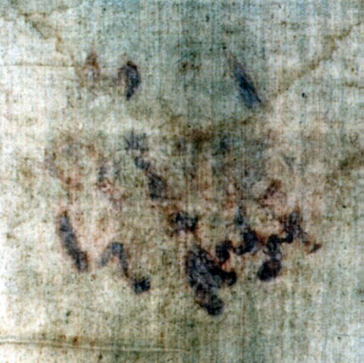
Figure 3: Image processing of Fig. 2 to evidence the bloodstains on the nape of the neck
The
peculiar angular profile of some bloodstains suggests the presence of objects
interposed between the back of the head and the HST. These objects would have
influenced the path of the blood flows in the occipital area. If the branches
were leafy, it is easy to identify these objects as leaves, as hypothesized by
O. Scheuermann in 1983 32. It can
thus be assumed that some leaves possibly of Rhamnus lycioides, see (Figure
4) remained tangled in the hair, due to adhesion to clots and that these
then interfered with the path of the blood fluid. For example, (Figure 5)
shows a possible interference between bloodstains on the HST and leaves.
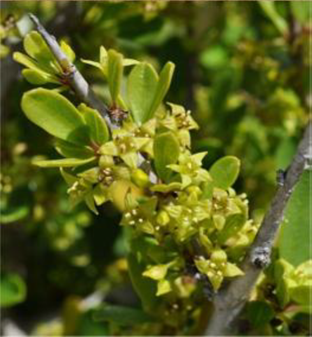
Figure
4: Leaves
of Rhamnus Lycioides (actaplantarum.org/forum/viewtopic.php?t=38880)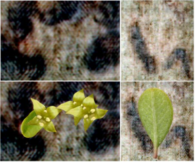
Figure
5: Possible
interference from objects interposed between the HST and the back of the head
At the top,
the peculiar angular profile of some bloodstains on the back of the head in the
post-processed image of (Figure 3) does not rule out the presence of
leaves. At the bottom, possible overlap of Rhamnus Lycioides leaves on these
bloodstains.
Finally, it
is worth noting the peculiar nature of many bloodstains on the nape of the
neck. They exhibit irregular circular shapes characterized by a light central
point and a darker outer contour, often serrated because they follow the linen
twill of the HST, see (Figure 6).
These
shapes suggest the possible presence of external corpuscles, such as small
stones or aromatic substances like myrrh (perhaps mixed with aloe), which are
also mentioned in the Gospel of John (19:39-40).
Unlike
other bloodstains present on the HST, those on the nape of the neck display two
distinct chromatic areas imprinted on the linen: a lighter rounded zone, which
appears to represent the imprint of the corpuscles in question, surrounded by a
darker outer area that frequently blends with the rest of the bloodstains.
These
circular shapes are present both in the more pronounced bloodstains, which can
be attributed to trickling that occurred during burial and in the fainter,
lighter ones, which are believed to have formed at an earlier moment.
We can
therefore think that direct contact between the cloth and hair soaked in blood
and ointments may have generated these particular formations. However, the
light, rounded imprints suggest the addition of corpuscles of a different
nature distributed almost uniformly across the entire posterior region of the
head, which somehow reduced or prevented the soaking up of blood onto the linen
of the HST.
To better
understand how these rounded shapes may have formed, attempts were made to
experimentally reproduce them starting from the bloodstains of the nape of the
neck.

Figure
6: Image
enhancing of bloodstains on the nape of the neck evidencing irregular circular
shapes characterized by a light central point and a darker outer contour. They
are both evident in both in the more pronounced bloodstains on the right and in
the fainter, lighter ones on the left
Experimental Tests and
Results
To better
interpret the peculiar bloodstains on the nape of the neck, which display
sharply defined edges, experimental tests were conducted using a red vinyl
fluid diluted with water.
The first
test aimed to verify whether the bloodstains could have resulted from a simple
dripping of blood through the hair under the sole influence of gravity. A
synthetic lock of hair was placed on a flat horizontal surface, with fabric
positioned beneath it. The red fluid was then allowed to fall freely from
above.
A partial
filtering effect by the hair was observed; however, the shape of the stain
produced on the fabric differed significantly from that of the actual
bloodstain. Specifically, the shapes and distribution of the stains obtained
did not match in terms of either quantity or directionality, thus ruling out
the hypothesis of simple vertical dripping.
The second
test was then performed to simulate the slow leakage of blood from a wound in
the occipital area, taking into account the contact force exerted by the weight
of the head on the sheet through the humid hair.
To do this,
the occipital region of the head was simulated using a PVC balloon, pierced in
the area of the nape with pinpoint cuts compatible with wounds caused by a
crown of thorns. The aqueous mixture of red vinyl glue was introduced into the
deflated balloon, which was then inflated, placed over a lock of hair and
finally pressed to simulate head-to-sheet contact, see (Figure 7).
The result,
shown in (Figure 8), evidenced drips that were less abundant as in the
first test and more regular compared to simple dripping.

Figure
7:
Experimental test performed simulating of the occipital region using a PVC
balloon. The balloon filled with red fluid (top) was then pierced (center) and,
pressurized, brought into contact with hair and fabric
The
third test attempted
to reproduce the observed well defined shapes, characterized by a central
unstained lighter area surrounded by an angular profile with a very
well-defined outline.
To conduct
this test, the hypothesis was based on the idea that these marks could have
been produced by leaves, placed between the hair and the cloth as suggested by
O. Scheuermann 32.
A leaf was
therefore placed between the hair and the fabric, as shown in (Figure 9).
Using the same procedure as the second test, with the addition of leaves
between the hair, the experiment yielded shapes morphologically very similar to
those observed on the HST, with angular edges and a central area without traces
of color.
In
agreement with O. Scheuermann 32,
this result is therefore not inconsistent with the hypothesis that plant
material was placed between the back of the head and the fabric on the HST and
it provides support to the hypothesis that plant materials contributed to the
formation of some traces on the HST.

Figure
8:
Simulated bloodstains on fabric during the second test

Figure
9:
Third test: on the top experimental setup, on the bottom shapes morphologically
very similar to those observed on the HST, with angular profile and a central
area without traces of color
The
fourth test,
perhaps the most significant one, was conducted to experimentally reproduce an
interesting characteristic observed on the HST. The bloodstains display small,
irregular, rounded shapes within them, with a dark border and lighter spots
inside, as highlighted in (Figure 6). These shapes are evident both in
the more pronounced bloodstains and in the fainter, lighter ones and are not
easy to reproduce.
Several
experiments were therefore carried out using various materials placed between
the hair and the cloth or positioned beneath it. In particular, small spheres
such as fishing lead weights and pebbles a few millimetres in diameter were
initially used, placed either between the support surface and the cloth under
the PVC ballon with hair simulating the human head or between the cloth and the
PVC balloon with hair. Tests with these materials placed between the support
flat surface and the cloth yielded negative results, while those simulating the
presence of rounded objects placed between the hair and the cloth appeared
promising.
Based on
these results, it was then hypothesized that, given the documented use of myrrh
as a substance employed in funeral rites, grains of natural myrrh might have
been placed beneath the head with hair, to better preserve the corpse.
In this
final configuration, after the experimental red fluid had dried, the result
obtained showed a remarkable resemblance to the pattern observed on the HST:
the stains appeared light in the centre with a dark border, matching both in
shape and size, as shown in (Figure 10).
It is
thought that myrrh, composed of essential oils, gum resins and phenolic
compounds, partially dissolved upon contact with the red fluid (and thus also
with contact of human blood), forming a viscous reddish-brown fluid similar to
the outlines of the marks detected on the HST.
Therefore,
to explain the dark border of the rounded stains, the following hypothesis is
proposed: during the burial, half of the HST was laid on the stone, upon which,
at least in the area corresponding to the head, several grains of myrrh-possibly
mixed with aloe for enhanced anti-putrefactive action-were placed. The corpse
was then laid down and then the second half of the HST was placed over it.

Figure
10:
Fourth test: red stains produced by grains of natural myrrh interposed between
hair and cloth
Conclusive Remarks
This study
has investigated the origin of the bloodstains present in the occipital region
of the body image of Jesus on the HST, based on both image processing and
experimental analysis. The multidisciplinary approach, conducted through
numerous experimental tests, sought to reproduce the particular bloodstains
visible at the back of the head.
It is
important to recall that the tests performed are based on models developed from
simplified hypotheses, which, while providing valid support, do not yet allow
for the absolute faithful reproduction of the complexity of the original
events. Nevertheless, many similarities were observed between the results
obtained from the experimental tests and the features observed in the analysed
areas of the HST.
The initial
experimental phases ruled out the possibility that simple vertical fluid flow
could explain the traces present on the HST. To achieve a more realistic and
consistent fluid distribution with the analysed image, a punctured balloon
containing pressurized red fluid was placed in contact with the fabric.
This
experimental setup allowed for the exploration and simulation of more complex
and realistic conditions, including the insertion of leaves between strands of
hair to simulate the angular yet well-defined profile of some bloodstains on
the HST. One may therefore hypothesize that the crown of thorns was made of
leafy branches, which left some leaves trapped between the hair, blood and
sweat after its removal.
The attempt
to reproduce the light stains with dark borders visible in the examined area of
the HST using solid objects, such as small weights and discs, led to the
hypothesis that granular myrrh was used, placed between the strands of hair and
the HST. Due to its physical properties, the myrrh produced patterns
compatible—both geometrically and chromatically-with those evident on the HST.
Given the
interesting results obtained, similar tests could be conducted in the future
using a cadaver and human blood. The work carried out thus provides a new
contribution to the analysis of the bloodstains on the HST in the occipital
region.
Acknowledgments
The authors
thank the Archdiocese of Torino for the HST photos made by G. Durante and Dr.
Gilbert Lavoie for Miller’s UV photos (https://shroudphotos.com/) Thanks also to Andrea Valmorbida who
participated in the experimental analysis.
Conflicts of Interest
The authors
declare no conflict of interest.
References
10. Fanti G. Open issues
regarding the Turin Shroud. Scientific Research and Essays 2012;7(29):2507.
13.
Garello E. The Shroud and the Popes, Corsi Ed. Turin
1984.
14.
Jackson J. The Shroud: A critical summary of data,
observations and hypotheses, Turin Shroud Center of Colorado, USA 2017.
15. Fanti G. Byzantine
coins influenced by the Shroud of Christ. Jenny Stanford Publishing Pte Ltd
2022.
18. McAvoy T. On
Radiocarbon Dating of the Shroud of Turin. Int J Archaeol 2021;9(2):34-44.
21. Phillips TJ. Shroud
irradiated with neutrons? Nature 1989;337.
31. Miller V. UV Photos of the Turin Shroud. © Vernon Miller 1978.
32.
Scheuermann, Oswald, Das Tuch, Verlag Friedrich
Pustet, Regensburg, Deutschland 1987.